






























Skin Tumor Excision
Skin Tumor Excision is a minor surgical procedure used to remove selected skin tumors or growths. Depending on the lesion, excision may be performed to treat a benign growth, relieve symptoms, establish a diagnosis, or remove a suspected or confirmed skin cancer.
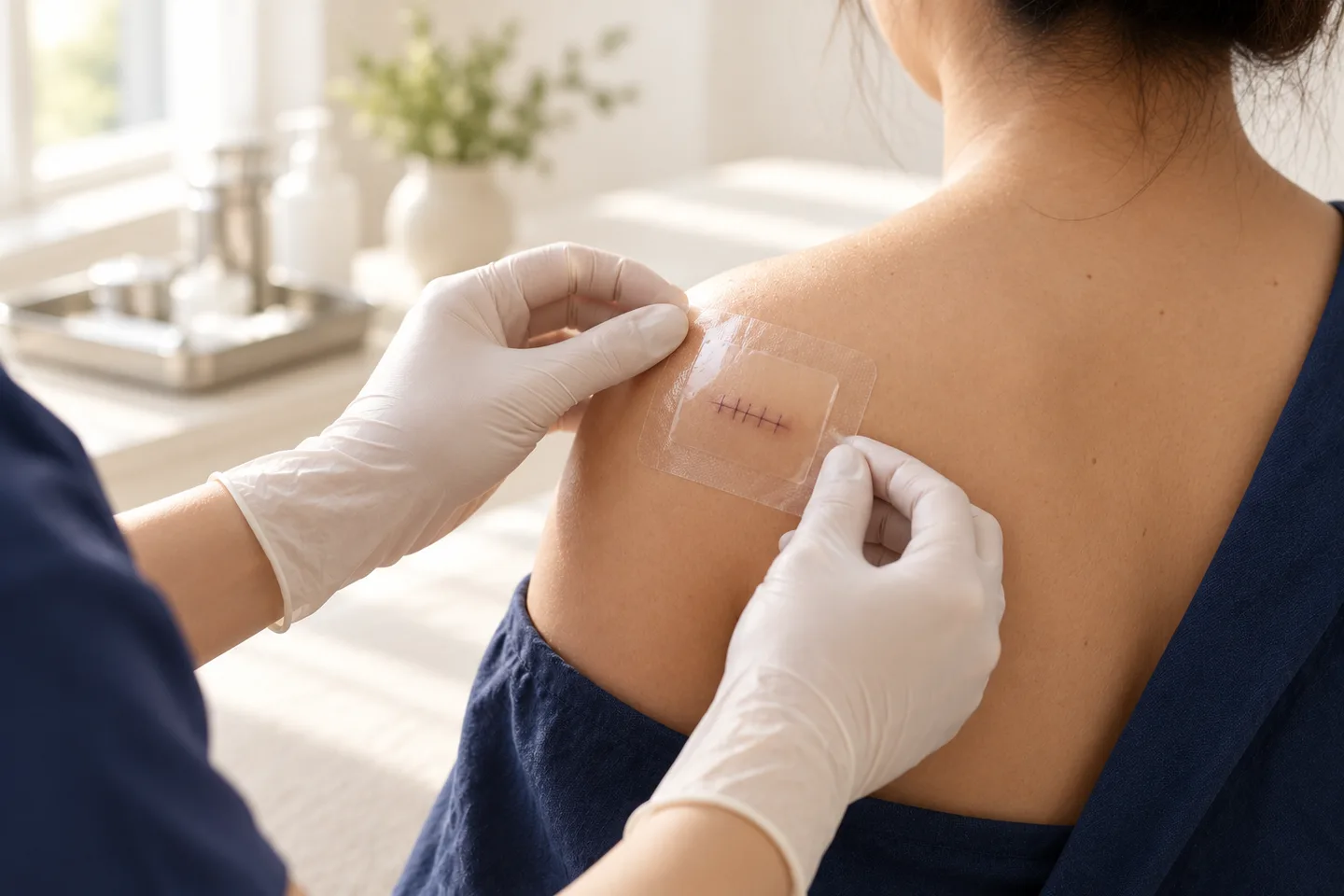
For many small and appropriately selected skin tumors, excision can be performed under local anesthesia. The lesion is removed with a scalpel, bleeding is controlled, the wound is usually closed with sutures, and the tissue is sent for Histopathology when appropriate. Histopathological examination is especially important when the diagnosis is uncertain or when a malignant or potentially malignant tumor is being evaluated.
The most important part of Skin Tumor Excision is not simply removing a visible lump. The clinician must first decide:
What is the lesion, does it need to be biopsied or completely removed, and what surgical approach is appropriate for that diagnosis?
A benign cyst, a suspicious mole, basal cell carcinoma, squamous cell carcinoma, and melanoma should not automatically be approached in the same way.
What Is a Skin Tumor?
A skin tumor is an abnormal growth of cells involving the skin or structures beneath it.
The word “tumor” does not automatically mean cancer.
Skin tumors can be broadly divided into:
Benign skin tumors
These are non-cancerous growths. Examples include:
- Lipoma
- Epidermoid Cyst
- Dermatofibroma
- Seborrheic Keratosis
- Skin Tag
- Some benign melanocytic nevi
- Pilomatricoma
- Other benign adnexal tumors
Premalignant or precancerous lesions
These have abnormal cells with the potential to progress or may represent an early stage in the pathway toward skin cancer.
Examples may include selected cases of:
- Actinic Keratosis
- Bowen’s Disease / Squamous Cell Carcinoma in Situ
Malignant skin tumors
These are skin cancers and include:
- basal cell carcinoma
- Squamous Cell Carcinoma
- Melanoma
- Less common cutaneous malignancies
The appropriate treatment depends on the exact diagnosis.
What Is Skin Tumor Excision?
Skin Tumor Excision means surgically cutting out a skin lesion.
When the entire lesion is removed primarily for diagnosis, the procedure may also be called an Excisional Biopsy.
Excision can have more than one purpose:
- Remove a benign tumor that is symptomatic or bothersome
- Remove a lesion for diagnostic confirmation
- Treat selected skin cancers
- Determine whether a tumor has been completely removed
- Obtain tissue for Histopathology
- Relieve pain, bleeding, irritation, or repeated inflammation
An Excisional Biopsy provides the pathologist with the complete lesion, which can be particularly useful when the overall architecture of the tumor is important for diagnosis.
Does Every Skin Tumor Need to Be Removed?
No.
Many benign skin tumors can safely be observed if they:
- Have a clear benign diagnosis
- Remain stable
- Cause no symptoms
- Are not repeatedly traumatized
- Do not bother the patient cosmetically
Removal may be considered when a lesion:
- Is painful
- Frequently becomes irritated
- Bleeds
- Catches on clothing
- Repeatedly becomes inflamed
- Is enlarging
- Causes functional problems
- Is cosmetically bothersome
- Has an uncertain diagnosis
- Has features suspicious for skin cancer
The decision should be based on diagnosis rather than simply whether a lesion is raised or visible.
Why Is Diagnosis Important Before Skin Tumor Excision?
Different tumors require different surgical strategies.
For example:
A benign superficial tumor may require only removal of the visible lesion.
A cyst usually needs its cyst wall removed to reduce the risk of recurrence.
A suspected skin cancer may require removal with an appropriate surgical margin.
A suspicious melanocytic lesion may require an Excisional Biopsy designed specifically to establish the diagnosis before definitive treatment margins are determined.
Therefore, performing a destructive procedure without first understanding what a lesion is can sometimes make subsequent diagnosis or treatment more difficult.
How Does a Dermatologist Evaluate a Skin Tumor?
Assessment usually begins with medical history and examination.
The dermatologist may ask:
- How long has the lesion been present?
- Has it changed recently?
- Is it growing?
- Has the color changed?
- Does it bleed?
- Does it itch or hurt?
- Has it ulcerated?
- Has it been treated previously?
- Has it returned after removal?
- Do you have a personal history of skin cancer?
- Do you have a family history of Melanoma or other skin cancers?
The examination may assess:
- Size
- Shape
- Color
- Border
- Symmetry
- Surface
- Consistency
- Depth
- Mobility
- Ulceration
- Bleeding
- Relationship to surrounding structures
For pigmented or suspicious lesions, Dermoscopy may provide additional diagnostic information.
Dermoscopy performed by an experienced clinician improves diagnostic assessment of melanocytic lesions and is recommended as part of melanoma evaluation.
What Warning Signs Should Be Evaluated Before Removal?
A skin lesion should be medically evaluated rather than simply cosmetically removed if it shows concerning features.
These may include:
- Rapid growth
- Recent significant change
- Irregular shape
- Asymmetry
- Multiple or changing colors
- Irregular borders
- Spontaneous bleeding
- Repeated crusting
- Persistent ulceration
- A wound that does not heal
- Increasing firmness
- Fixation to deeper tissue
- New persistent pain
- Recurrence after previous removal
- A lesion that looks different from the patient’s other moles or growths
A change, itch, or bleeding can be among the reasons a dermatologist may consider a lesion suspicious enough to biopsy.
Benign Skin Tumor vs Skin Cancer
It is not always possible for a patient to determine this from appearance alone.
| More typical of a benign lesion | Features requiring closer evaluation |
|---|---|
| Stable over a long period | Rapid or progressive growth |
| Symmetrical | New asymmetry |
| Uniform appearance | Irregular or changing pigmentation |
| Smooth border | Irregular border |
| No bleeding | Spontaneous bleeding |
| No ulceration | Persistent ulceration |
| Soft or mobile where expected | Firm or fixed |
| No major recent change | Significant recent change |
These features are guides rather than diagnostic rules.
Some skin cancers can look harmless, while some benign lesions can appear unusual.
What Types of Skin Tumors Can Be Removed Surgically?
Excision may be used for many different lesions, depending on diagnosis, size, depth, and location.
Examples include:
- Lipoma
- Epidermoid Cyst
- Dermatofibroma
- Selected benign nevi
- Pilomatricoma
- Selected adnexal tumors
- Recurrent or diagnostically uncertain lesions
- basal cell carcinoma
- Squamous Cell Carcinoma
- Squamous Cell Carcinoma in Situ
- Selected Melanoma lesions as part of the diagnostic or definitive surgical pathway
Not every lesion should be treated by standard excision, and some lesions are better managed with another technique.
What Is an Excisional Biopsy?
An Excisional Biopsy removes the entire visible lesion so it can be examined by a pathologist.
It can therefore be both:
Diagnostic — determining what the tumor is.
and
Therapeutic — removing the lesion.
This approach is particularly valuable when examination of the entire lesion and its architecture is important for diagnosis.
Excisional Biopsy vs Incisional Biopsy
These procedures serve different purposes.
Excisional Biopsy
The entire lesion is removed.
It may be appropriate for relatively small lesions when complete removal can be performed safely.
Incisional Biopsy
Only part of the lesion is removed for diagnosis.
It may be appropriate when:
- The tumor is large
- Complete initial removal would produce an unnecessarily large wound
- The lesion is in a complex location
- The diagnosis needs to be established before definitive surgery
- A deeper process needs to be sampled
An Incisional Biopsy usually establishes the diagnosis rather than serving as definitive treatment.
Excision vs Punch Biopsy
A Punch Biopsy removes a small cylindrical sample of skin.
It is useful for many dermatological diagnoses and selected tumors.
However, a small partial sample is not appropriate for every lesion.
For a lesion suspicious for Melanoma, complete full-thickness excision with a narrow margin is generally preferred when technically feasible because the pathologist needs appropriate tissue to assess important features such as tumor depth.
What Is Histopathology?
Histopathology is microscopic examination of tissue after it has been removed.
The excised specimen is processed in a laboratory and examined by a pathologist or Dermatopathologist.
Histopathology can help determine:
- What type of tumor it is
- Whether it is benign or malignant
- The subtype of the tumor
- Important microscopic features
- Whether further treatment is necessary
- Whether tumor reaches the edge of the excised specimen
For suspected skin cancer, microscopic examination is required to establish the diagnosis.
Why Should an Excised Skin Tumor Be Sent for Histopathology?
A lesion can sometimes look clinically benign but reveal a different diagnosis under the microscope.
Histopathology is particularly important when:
- The diagnosis is uncertain
- The lesion is changing
- Skin cancer is suspected
- The lesion has returned after previous treatment
- The tumor has unusual features
- Surgical margins need assessment
Surgically removed skin lesions should be submitted for pathological examination.
What Is a Surgical Margin?
A Surgical Margin is the edge of apparently normal tissue removed around or beneath a tumor.
Margins are particularly important in skin cancer surgery.
After excision, the pathologist may assess whether tumor cells extend to the edge of the specimen.
A report may describe the margin as clear or involved, although terminology varies between laboratories.
Margin status can affect whether additional treatment is needed.
Does Every Skin Tumor Need a Margin?
No.
The required approach depends on the diagnosis.
A clearly benign lesion does not require the same oncologic surgical margin as a malignant tumor.
For skin cancers, the appropriate margin depends on factors such as:
- Tumor type
- Tumor size
- Tumor location
- Histological subtype
- Tumor borders
- Whether it is primary or recurrent
- Other high-risk features
Therefore, there is no single surgical margin that should be used for every skin tumor.
Skin Tumor Excision for Suspected Melanoma
A suspicious pigmented lesion requires particularly careful planning.
When Melanoma is suspected and the lesion can be completely removed safely, guidelines generally favor a full-thickness complete Excisional Biopsy with a narrow clinical margin to establish the diagnosis.
If Melanoma is confirmed, definitive wider excision is subsequently planned according to pathological factors, especially the measured tumor thickness.
This is why performing a large definitive excision before establishing the melanoma diagnosis is usually not the initial approach.
Skin Tumor Excision for basal cell carcinoma
basal cell carcinoma is a common form of skin cancer.
Standard Surgical Excision can be appropriate for many lesions, but management depends on:
- Location
- Size
- Clinical borders
- Histological subtype
- Whether the tumor is recurrent
- Other risk factors
Some higher-risk tumors or tumors in anatomically important areas may be better suited to techniques that provide more complete margin assessment, such as Mohs Micrographic Surgery.
Skin Tumor Excision for Squamous Cell Carcinoma
Squamous Cell Carcinoma is another form of skin cancer that commonly requires surgical treatment.
The surgical plan depends on factors such as:
- Size
- Location
- Depth
- Histological differentiation
- Perineural involvement
- Immune status
- Whether the tumor is recurrent
- Other high-risk pathological features
Higher-risk tumors may require wider management than routine Minor Skin Surgery.
What Is Mohs Micrographic Surgery?
Mohs Micrographic Surgery is a specialized skin cancer surgery that removes the tumor in stages while examining the surgical margins microscopically during the procedure.
It is particularly valuable in selected skin cancers when:
- Tissue preservation is important
- The tumor is in a high-risk anatomical area
- Borders are difficult to define
- The tumor has returned
- Histology indicates higher risk
- Complete margin assessment is especially valuable
Mohs Surgery examines the surgical margin more comprehensively than conventional excision and can preserve more uninvolved tissue in appropriate cases.
Not every skin cancer requires Mohs Surgery.
Standard Excision vs Mohs Surgery
| Standard Surgical Excision | Mohs Micrographic Surgery |
|---|---|
| Tumor removed in one procedure | Tumor removed in stages |
| Specimen sent for conventional Histopathology | Margins examined during the procedure |
| Appropriate for many skin tumors | Selected mainly for particular skin cancers |
| Pathology result usually follows later | Margin assessment occurs during surgery |
| Appropriate margins selected before excision | Additional tissue removed only where residual tumor is identified |
The correct choice depends on tumor type and risk rather than simply patient preference.
How Is Skin Tumor Excision Performed?
The exact technique varies according to the tumor.
A typical Minor Skin Surgery procedure may include the following steps.
1. Confirm the Surgical Site
The dermatologist identifies the exact lesion and reviews the diagnosis or reason for removal.
This is especially important when multiple similar lesions are present.
2. Mark the Lesion and Planned Excision
The lesion and planned incision are marked.
For some tumors, an appropriate surgical margin is also marked.
3. Clean the Skin
The surgical area is cleaned with an antiseptic solution and prepared for surgery.
- Inject Local Anesthesia
Local anesthetic is injected around the lesion.
The injection may cause brief stinging or burning.
Once the area is adequately numb, the patient should generally feel pressure or movement rather than sharp pain.
Local anesthesia is commonly used for cutaneous excisional procedures.
5. Excise the Tumor
The dermatologist uses a scalpel to remove the lesion at the planned depth and margin.
Many excisions are designed in an elliptical or fusiform shape because this allows the wound edges to be closed in a controlled manner.
6. Control Bleeding
Bleeding is controlled before wound closure.
7. Close the Wound
Depending on wound depth and tension, closure may involve:
- Superficial sutures
- Buried absorbable sutures
- Layered closure
Deep sutures can reduce tension and help close deeper spaces beneath the skin.
8. Apply a Dressing
The wound is cleaned and covered.
9. Send Tissue for Histopathology
When indicated, the specimen is labeled and sent to the pathology laboratory.
Why Is the Excision Often Elliptical?
A round tumor is often removed through an elliptical or fusiform surgical design.
This may seem counterintuitive to patients because the final scar can be longer than the diameter of the original lesion.
The reason is that an elliptical design often allows the skin edges to come together more smoothly and reduces bunching of tissue at the ends of the wound.
The incision may also be aligned with natural skin tension lines when possible to improve wound healing and scar orientation.
Why Can the Scar Be Longer Than the Tumor?
This is an important expectation to discuss before surgery.
Removing a 1-centimeter lesion does not necessarily produce a 1-centimeter scar.
To close the resulting defect smoothly, the incision often extends beyond the visible lesion.
Additional factors include:
- Required Surgical Margin
- Tumor depth
- Skin tension
- Anatomical location
- Need for appropriate wound closure
Therefore, the final scar can be several times longer than the visible diameter of the tumor.
Does Skin Tumor Excision Hurt?
The most noticeable discomfort is usually the injection of the local anesthetic.
Patients may temporarily feel:
- Stinging
- Burning
- Pressure
Once adequate anesthesia has been achieved, patients may still notice pulling, touching, or movement but should not normally feel sharp surgical pain.
After the anesthetic wears off, some soreness or tenderness is expected.
How Long Does Skin Tumor Excision Take?
There is no universal procedure time.
A small superficial lesion may be relatively straightforward.
A larger or deeper tumor may require additional time for:
- Careful dissection
- Bleeding control
- Removal with appropriate margins
- Layered closure
- Reconstruction
The anatomical location can matter as much as the lesion size.
How Is the Wound Closed?
Several techniques may be used.
Primary Closure
The two wound edges are brought together and sutured.
This is commonly used for smaller excisions.
Layered Closure
Deep absorbable sutures are used beneath the skin, followed by superficial closure.
This is useful when the wound is deeper or under greater tension.
Secondary Intention Healing
In selected situations, a wound may be left to heal naturally from the base upward.
Skin Flap
Nearby skin is moved to cover a defect.
Skin Graft
Skin is taken from another location to cover the surgical wound.
Larger excisions may require a flap or graft rather than simple direct closure.
Will Skin Tumor Excision Leave a Scar?
Yes.
Complete surgical removal of a skin lesion necessarily produces some degree of scarring.
The final appearance depends on:
- Size of the excision
- Location
- Surgical Margin
- Wound tension
- Direction of the scar
- Surgical technique
- Infection or wound complications
- Individual healing characteristics
- History of Keloid
- History of Hypertrophic Scar
- Sun exposure during healing
Scars continue to change for months after surgery.
Can Skin Tumor Excision Be Scarless?
No surgical procedure that cuts through the skin can guarantee no scar.
A more realistic goal is to:
- Remove the lesion appropriately
- Preserve healthy tissue where possible
- Position the scar thoughtfully
- Reduce wound tension
- Prevent complications
- Provide appropriate scar care during healing
For suspected or confirmed cancer, complete tumor management takes priority over making the shortest possible scar.
Skin Tumor Excision in Asian and Darker Skin
Skin surgery can be safely performed in people with Asian and darker skin tones, but postoperative pigment and scar changes deserve consideration.
Possible changes include:
- Post-Inflammatory Hyperpigmentation or PIH
- Hypopigmentation
- Persistent redness
- Hypertrophic Scar
- Keloid
Patients with a previous history of thick or spreading scars should mention this before surgery.
Scar risk also varies substantially by body site. Shoulders, upper chest, upper back, and some other high-tension areas may be more prone to prominent scarring.
What Are the Risks of Skin Tumor Excision?
Possible complications include:
- Pain
- Bleeding
- Bruising
- Swelling
- Hematoma
- Infection
- Wound separation
- Delayed healing
- Scar
- PIH
- Hypopigmentation
- Hypertrophic Scar
- Keloid
- Temporary numbness
- Persistent numbness
- Nerve injury
- Injury to nearby structures
- Reaction to dressings or adhesives
- Incomplete tumor removal
- Positive Surgical Margin
- Recurrence
The likelihood and significance of these complications depend heavily on the type, size, depth, and location of the tumor.
Can Skin Tumor Excision Cause Numbness?
Yes.
Small sensory nerves travel through the skin and subcutaneous tissue.
Temporary numbness near a surgical scar can occur because of swelling, stretching, or interruption of small nerve branches.
More significant nerve injury is less common but becomes more relevant when a tumor is:
- Deep
- Large
- Close to an important sensory nerve
- Close to a motor nerve
- In an anatomically complex location
This is one reason some tumors may require referral rather than routine office-based excision.
Can a Skin Tumor Come Back After Excision?
Yes, depending on the tumor.
Recurrence may occur because:
- Part of the lesion remains
- Tumor extends beyond the apparent clinical border
- A cyst wall was incompletely removed
- The biological behavior of the tumor predisposes to recurrence
- A malignant tumor has involved margins
- A new lesion develops near the previous site
A recurrent lesion should be reassessed rather than automatically treated in the same way again.
What Does a Positive Surgical Margin Mean?
A positive or involved margin generally means tumor cells are present at the edge of the specimen examined by the pathologist.
This may indicate that some tumor could remain in the patient.
What happens next depends on:
- Tumor type
- Which margin is involved
- Tumor risk level
- Anatomical location
- Previous treatments
- Patient factors
Options may include additional excision, Mohs Surgery, another form of treatment, or selected observation depending on the diagnosis.
Margin interpretation is especially important for malignant tumors.
What Happens After the Pathology Result?
The next step depends on the diagnosis.
Benign tumor completely removed
No further treatment may be required apart from wound and scar care.
Benign tumor but incomplete removal
Observation or further treatment may be discussed depending on symptoms and recurrence risk.
Precancerous lesion
Additional treatment may or may not be necessary depending on diagnosis and margin status.
Skin cancer with adequate margins
Follow-up and future skin surveillance may be recommended according to the cancer type and risk.
Skin cancer with involved or inadequate margins
Additional surgery or another form of treatment may be necessary.
Unexpected or unusual tumor
Additional pathology tests, imaging, wider excision, or referral to another specialist may be needed.
Recovery After Skin Tumor Excision
Recovery varies considerably.
A small lesion removed from a low-tension area may cause little disruption.
A larger operation on the back, shoulder, leg, or near a joint can require greater activity modification.
During the first few days, patients may notice:
- Tenderness
- Mild swelling
- Bruising
- Tightness around the sutures
- Small amounts of spotting on the dressing
Symptoms should generally improve rather than progressively worsen.
Wound Care After Skin Tumor Excision
Follow the instructions provided by the treating clinician because wound care varies according to the procedure.
General principles include:
- Keep the initial dressing in place as instructed
- Keep the wound clean
- Wash gently when permitted
- Avoid picking at crusts or sutures
- Avoid unnecessary friction
- Avoid excessive pulling on the wound
- Attend wound checks when advised
- Have sutures removed at the appropriate time
- Protect the healing scar from excessive sun exposure
- Watch for infection
Dermatologic surgery guidance emphasizes appropriate dressing, gentle cleansing, and protection of the wound during healing.
When Are Sutures Removed?
The timing depends on:
- Anatomical location
- Wound tension
- Size of the operation
- Type of suture
- Healing progress
Facial sutures are generally removed sooner than sutures on the trunk or lower limbs.
Some deeper sutures are absorbable and remain beneath the skin.
There is no single removal day appropriate for all Skin Tumor Excision procedures.
When Can I Exercise After Skin Tumor Excision?
This depends on the location and size of the wound.
Exercise can increase wound tension, which may contribute to:
- Bleeding
- Swelling
- Wound separation
- Wider scars
Activities involving the operated area may need temporary restriction.
For example, an excision over the shoulder or upper back can be affected by weight training and arm movement more than a small excision in a low-tension area.
When Can I Swim?
Swimming should generally wait until the wound has sealed adequately and the treating clinician considers it safe.
A recently sutured wound should not be subjected to prolonged soaking or unnecessary contamination.
This is particularly relevant for tourists planning beach, pool, or island travel after Minor Skin Surgery in Thailand.
Can I Fly After Skin Tumor Excision?
Flying is often possible after uncomplicated Minor Skin Surgery, but the question should be considered in the context of postoperative care.
International patients should plan for:
- Wound checks
- Dressing changes
- Suture removal
- Histopathology results
- Possible further excision
- Management of complications if they arise
If a tumor is being removed because skin cancer is suspected, leaving Bangkok before the pathology result is available may complicate further treatment planning.
Warning Signs After Surgery
Contact your clinician if you develop:
- Increasing pain
- Progressive redness
- Increasing warmth
- Pus
- Fever
- Significant swelling
- Persistent bleeding
- Wound separation
- Rapidly enlarging swelling beneath the wound
- New significant weakness
- Severe or progressive numbness
- Darkening or loss of blood supply to the wound edges
- Unexpected deterioration
Some bruising, swelling, and mild discomfort can be normal. Progressive symptoms are more concerning.
When Should You See a Dermatologist for a Skin Tumor?
Consider a dermatology assessment when you notice:
- A new persistent growth
- A mole that is changing
- A lesion that repeatedly bleeds
- A non-healing sore
- A rapidly enlarging lump
- A recurrent lesion
- A painful or ulcerated growth
- An unusual pigmented lesion
- A firm or fixed tumor
- A lesion whose diagnosis is uncertain
Do not assume that every visible growth needs removal, and do not assume that every painless lesion is harmless.
What to Expect During a Skin Tumor Consultation in Bangkok
A useful consultation should establish the diagnosis and treatment pathway before surgery.
Important questions include:
What is the likely diagnosis?
The clinician should distinguish a benign tumor from a lesion that requires Biopsy or cancer evaluation.
Is Dermoscopy needed?
Dermoscopy may be especially useful for pigmented and selected non-pigmented lesions.
Should the lesion be biopsied or completely excised?
Some tumors are suitable for Excisional Biopsy, while others may require a partial Biopsy first.
What Surgical Margin is appropriate?
The answer depends on whether the lesion is benign, suspicious, or confirmed malignant.
Can the procedure be done using local anesthesia?
Many small superficial tumors can be treated this way, but deeper or complex tumors may require a different setting.
What will the scar look like?
The likely scar length, orientation, and risk of Keloid or Hypertrophic Scar should be discussed.
Will the specimen be sent for Histopathology?
This is particularly important for tumors that are uncertain or potentially malignant.
When will the pathology result be available?
Patients should understand how and when they will receive the result.
Could further surgery be required?
If Histopathology identifies malignancy or involved margins, additional treatment may be necessary.
Skin Tumor Excision for Tourists and International Patients in Bangkok
International patients should plan Minor Skin Surgery around the entire care pathway, not just the procedure date.
Before scheduling Skin Tumor Excision in Bangkok, consider:
- Whether diagnosis is clear before surgery
- Whether a Biopsy is required first
- Whether the lesion can be completely removed safely
- Whether Histopathology will be performed
- How pathology results will be communicated
- Whether further surgery could be required
- When sutures need removal
- How long wound care is required
- When swimming can resume
- Whether planned flights affect follow-up
- What to do if infection or bleeding develops after leaving Thailand
For a suspicious skin tumor, the safest plan may require more than one visit.
Frequently asked questions
What is Skin Tumor Excision?
It is surgical removal of a skin tumor through an incision, often with the tissue subsequently examined by Histopathology.
Is Skin Tumor Excision considered Minor Skin Surgery?
Many small superficial tumors can be removed as Minor Skin Surgery under local anesthesia. Large, deep, complex, or high-risk malignant tumors may require a different surgical setting.
Does every skin tumor need excision?
No. Many clearly benign, asymptomatic tumors can be observed.
Can a dermatologist tell if a tumor is benign just by looking?
Many lesions have characteristic clinical appearances, and Dermoscopy can improve assessment. However, Histopathology may be required when the diagnosis is uncertain or skin cancer is suspected.
What is the difference between a skin tumor and skin cancer?
A skin tumor simply means an abnormal tissue growth. It can be benign, precancerous, or malignant.
What skin tumors can be surgically removed?
Examples include Lipoma, Epidermoid Cyst, Dermatofibroma, selected nevi, adnexal tumors, basal cell carcinoma, Squamous Cell Carcinoma, and other selected tumors.
What is an Excisional Biopsy?
It is complete removal of a lesion primarily so the entire specimen can be examined microscopically.
What is the difference between Excisional Biopsy and Incisional Biopsy?
An Excisional Biopsy removes the whole lesion. An Incisional Biopsy removes only part of it for diagnosis.
What is Histopathology?
Histopathology is microscopic examination of removed tissue by a pathologist.
Should every excised skin tumor be sent for Histopathology?
Pathological examination is particularly important when diagnosis is uncertain, the lesion has unusual features, or malignancy is possible.
What is a Surgical Margin?
It is the edge of tissue removed around a tumor. Margin assessment is especially important in skin cancer surgery.
What does a clear margin mean?
It generally means tumor is not seen extending to the examined edge of the specimen.
What does a positive margin mean?
It means tumor cells are present at an examined edge of the specimen and additional management may be required.
Can a suspicious mole simply be shaved off?
Not always. If Melanoma is suspected, obtaining an appropriate full-thickness specimen is important for diagnosis and treatment planning.
How is suspected Melanoma biopsied?
When feasible, complete full-thickness Excisional Biopsy with a narrow clinical margin is generally preferred to establish the diagnosis.
Can basal cell carcinoma be treated with Surgical Excision?
Yes, standard Surgical Excision is appropriate for many basal cell carcinomas, although some higher-risk tumors may require another approach such as Mohs Surgery.
Can Squamous Cell Carcinoma be treated with excision?
Yes. Surgical treatment is commonly used, but the approach depends on tumor risk factors.
What is Mohs Surgery?
Mohs Micrographic Surgery removes skin cancer in stages while examining the surgical margins during the procedure.
Does Skin Tumor Excision hurt?
The local anesthetic injection can sting briefly. After the area is numb, sharp surgical pain should generally not be felt.
Can Skin Tumor Excision be performed with local anesthesia?
Many small superficial tumors can be removed using local anesthesia.
How long does the procedure take?
It varies according to tumor size, depth, location, required margin, and wound reconstruction.
Why is the scar longer than the tumor?
An elliptical incision is often used to allow the skin edges to close smoothly, so the final scar may be longer than the visible lesion.
Does Skin Tumor Excision leave a scar?
Yes. Complete surgical excision necessarily leaves a scar.
Can Skin Tumor Excision be scarless?
No surgical removal through an incision can guarantee no scar.
Can scars become Keloid?
Yes, particularly in predisposed individuals and certain anatomical areas.
Can surgery cause PIH in Asian skin?
Yes. Post-Inflammatory Hyperpigmentation can occur around healing surgical wounds, particularly in more pigmented skin.
Can a skin tumor come back after surgery?
Yes. Recurrence depends on the tumor type, completeness of removal, biological behavior, and other factors.
Can I exercise after Skin Tumor Excision?
Usually eventually, but strenuous activity may need to be temporarily restricted depending on wound location and tension.
Can I swim after surgery?
Swimming should generally wait until the wound has healed sufficiently.
Can I fly after Skin Tumor Excision?
Often yes after uncomplicated minor surgery, but wound follow-up, suture removal, and pathology results should be considered.
How long do sutures remain?
Timing depends on wound location, tension, and the type of suture used.
What should I do if the wound becomes red and painful?
Progressive redness, warmth, pain, swelling, pus, or fever should prompt medical review.
What happens if Histopathology unexpectedly shows skin cancer?
The pathology result is reviewed to determine the tumor type, margins, and other relevant features. Additional excision, Mohs Surgery, surveillance, or specialist management may be recommended depending on the diagnosis.
Can tourists have Skin Tumor Excision in Bangkok?
Yes when clinically appropriate, but international patients should make sure there is enough time for wound care, Histopathology results, suture removal, and any additional treatment that may be required.
Related conditions
-
Skin Biopsy (การวินิจฉัย / Diagnosis) Skin Biopsy (การวินิจฉัย / Diagnosis) A skin biopsy is a minor medical procedure in which a dermatologist removes a small sample of skin so it can be examined under a microscope. It is one of the most important diagnostic tools in dermatology because many skin conditions cannot be confirmed by appearance alone.

-
Excisional Biopsy Excisional Biopsy An excisional biopsy means completely cutting out a skin lesion, usually through the full thickness of the skin. The removed tissue is then examined under a microscope.

-
Histopathology Histopathology Histopathology is the microscopic examination of tissue to identify disease. In dermatology, this usually means examining a piece of skin removed during a skin biopsy or surgical excision.

-
Dermoscopy Dermoscopy Dermoscopy is a non-invasive examination technique that allows a dermatologist to inspect skin lesions with greater detail than is possible with the naked eye alone.
