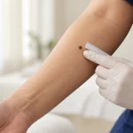
Skin Biopsy (การวินิจฉัย / Diagnosis)
A skin biopsy is a minor medical procedure in which a dermatologist removes a small sample of skin so it can be examined under a microscope. It is one of the most important diagnostic tools in dermatology because many skin conditions cannot be confirmed by appearance alone.
A skin biopsy may be recommended for a suspicious mole, a possible skin cancer, a persistent rash, an unusual growth, certain infections, blistering diseases or other skin conditions where microscopic examination can provide a more accurate diagnosis.
For patients in Bangkok, including Thai patients, expatriates, tourists and international patients, understanding why a biopsy is recommended—and what happens afterwards—can make the process much less worrying.
What Is a Skin Biopsy?
A skin biopsy means removing a small amount of skin or an entire skin lesion for laboratory examination.
The tissue is usually sent to a pathologist or dermatopathologist, a doctor trained to diagnose diseases by examining tissue under a microscope. The pathology report can help determine what the lesion is and whether further treatment is required.
A biopsy is not the same as simply removing a skin lesion for cosmetic reasons. Its primary purpose is usually diagnosis.
Does Having a Skin Biopsy Mean the Doctor Thinks You Have Cancer?
No.
Skin biopsies are commonly performed to investigate possible skin cancer, but they are also used to diagnose many non-cancerous conditions.
Examples include:
- Inflammatory rashes
- Psoriasis
- Blistering skin diseases
- Certain infections
- Unusual moles
- Benign skin growths
- Actinic keratosis
- Autoimmune skin disorders
- Hair and scalp disorders in selected cases
A dermatologist recommending a biopsy is often saying:
“The appearance alone is not enough to make the diagnosis confidently.
”
It does not automatically mean cancer is likely.
Why Is a Skin Biopsy Important for Suspected Skin Cancer?
If a lesion might be skin cancer, microscopic examination of tissue is generally required to confirm the diagnosis.
Visual examination and dermoscopy can provide important clues, but the tissue sample allows a pathologist to determine whether cancer cells are present and, when cancer is found, identify the type.
Possible diagnoses include:
- Basal cell carcinoma
- Cutaneous squamous cell carcinoma
- Melanoma
- Carcinoma in situ
- Benign mole
- Atypical mole
- Seborrheic keratosis
- Other benign or malignant skin conditions
Does Every Mole or Skin Growth Need a Biopsy?
No.
Most moles and skin growths do not need biopsy.
Dermatologists first assess factors such as:
- Clinical appearance
- Change over time
- Color
- Border
- Shape
- Size
- Surface
- Bleeding or ulceration
- Dermoscopic features
- Patient age
- Location
- Personal and family history of skin cancer
Clinical recommendations note that not every spot, mole or growth requires biopsy; biopsy is generally recommended when examination reveals features that warrant tissue diagnosis.
When Might a Dermatologist Recommend a Skin Biopsy?
A biopsy may be considered if you have:
- A new suspicious mole
- A mole that is changing
- An unusual pigmented lesion
- A non-healing sore
- A lesion that repeatedly bleeds or crusts
- A persistent pink or red patch
- A rapidly growing skin growth
- A possible basal cell carcinoma
- A possible squamous cell carcinoma
- A possible melanoma
- An unexplained chronic rash
- Blistering disease
- A skin condition that has not responded as expected to treatment
- An unusual lesion where the diagnosis remains uncertain
The decision should be based on the clinical question the biopsy needs to answer.
What Are the Main Types of Skin Biopsy?
There is no single best biopsy technique for every skin problem.
The dermatologist chooses the method according to:
- Suspected diagnosis
- Depth of the disease
- Size of the lesion
- Location
- Whether melanoma is a concern
- Whether the whole lesion should be removed
- Cosmetic considerations
- Need for sutures
Common techniques include:
- Shave biopsy
- Punch biopsy
- Incisional biopsy
- Excisional biopsy
- Curettage in selected lesions
- Snip biopsy for certain protruding lesions
Clinical guidelines describe these as common approaches used according to the depth and type of lesion being investigated.
Shave Biopsy
A shave biopsy removes a layer of tissue from the surface of the lesion using a blade.
It is often useful for relatively superficial lesions.
Examples may include certain:
- Seborrheic keratoses
- Basal cell carcinomas
- Superficial keratinizing lesions
- Raised benign lesions
- Other superficial growths
The main advantage is that it is usually quick and often does not require stitches.
The important limitation is depth. A superficial shave may not sample the full thickness of a deeper lesion. Standard medical literature notes that this can make evaluation of invasive disease difficult if the sample is not deep enough.
Will a Shave Biopsy Leave a Scar?
Usually yes, although the scar may be relatively subtle.
Possible appearances include:
- A lighter patch
- A darker patch
- A shallow depression
- A flat scar
The final appearance varies according to body site, depth of biopsy, skin type and individual healing.
Deep Shave or Saucerization Biopsy
A deeper form of shave biopsy removes more of the dermis than a superficial shave.
It can sometimes be used for selected pigmented lesions where a deeper sample is needed.
The depth is important when melanoma is part of the differential diagnosis because an inadequate sample can make accurate measurement of tumor depth difficult.
For this reason, biopsy technique for a suspected melanoma should be chosen carefully rather than using the same approach for every mole.
Punch Biopsy
A punch biopsy uses a small circular instrument that removes a cylindrical piece of skin.
Unlike a superficial shave, a punch biopsy can include:
- Epidermis
- Dermis
- Sometimes superficial subcutaneous tissue
This makes it useful when the dermatologist needs to evaluate deeper layers of the skin.
Common indications may include:
- Inflammatory skin diseases
- Some rashes
- Autoimmune skin disorders
- Suspected deeper tumors
- Selected pigmented lesions
- Scalp disorders
- Certain infections
Depending on punch size and location, one or more stitches may be required.
Incisional Biopsy
An incisional biopsy removes only part of a larger lesion.
It may be appropriate when:
- The lesion is too large to remove completely during diagnostic biopsy
- Only a representative area is needed
- The diagnosis will determine how definitive surgery should be planned
The biopsy site should be selected carefully because sampling the wrong part of a large or heterogeneous lesion can occasionally give incomplete information.
Excisional Biopsy
An excisional biopsy removes the entire visible lesion, usually with a scalpel.
The wound is generally closed with stitches.
This approach may be appropriate for selected lesions when:
- The lesion is relatively small
- Full-thickness tissue is important
- The entire lesion can be safely removed
- Melanoma is suspected and complete diagnostic sampling is appropriate
An excisional biopsy can be both diagnostic and remove the visible lesion, but it does not automatically mean that definitive cancer treatment is complete.
If pathology shows cancer, further surgery may still be required depending on:
- Diagnosis
- Margins
- Tumor depth
- Tumor type
- Risk features
- Treatment guidelines
What Type of Biopsy Is Best for Suspected Melanoma?
The goal is to obtain enough tissue for accurate diagnosis and assessment of important pathological features.
Clinical recommendations emphasize that a suspected melanoma biopsy needs to include enough of the lesion to allow accurate diagnosis, and inadequate depth can interfere with measuring how deeply melanoma has invaded.
When technically feasible, the dermatologist generally aims to obtain an appropriate full-thickness sample of the suspicious lesion.
However, the exact approach may need modification for:
- Very large lesions
- Facial lesions
- Nail lesions
- Acral lesions
- Difficult anatomical locations
Therefore, the most appropriate biopsy technique depends on the individual lesion rather than a single rule.
What Type of Biopsy Is Used for Basal Cell Carcinoma?
Several techniques can be appropriate.
Depending on the appearance and location, a dermatologist may use:
- Shave biopsy
- Punch biopsy
- Incisional biopsy
- Excisional biopsy in selected cases
Basal cell carcinoma can resemble other conditions, so a tissue sample may be needed before choosing treatment.
What Type of Biopsy Is Used for Squamous Cell Carcinoma?
The biopsy must be deep enough to provide useful information about the lesion.
Possible approaches include:
- Shave biopsy
- Punch biopsy
- Incisional biopsy
- Excisional biopsy
The best technique depends on the thickness, location and clinical appearance of the lesion.
Can Skin Biopsy Diagnose a Rash?
Yes.
Biopsy is not only used for tumors.
It may help distinguish between conditions that can look similar clinically, such as:
- Psoriasis
- Eczema-like inflammatory disorders
- Autoimmune diseases
- Vasculitis
- Drug reactions
- Blistering disorders
- Some infections
- Certain cutaneous lymphomas
Sometimes one biopsy is enough.
In other cases, more than one sample or a special laboratory study may be necessary.
Can Skin Biopsy Diagnose Infection?
Sometimes.
Microscopic examination may reveal evidence of:
- Fungal infection
- Bacterial infection
- Viral changes
- Parasitic disease
However, biopsy is not always the best first test.
Depending on the suspected infection, the dermatologist may instead or additionally perform:
- Skin scraping
- Potassium hydroxide examination
- Bacterial culture
- Fungal culture
- Molecular testing
- Special tissue stains
The diagnostic method depends on the suspected organism.
What Happens Before a Skin Biopsy?
Before the procedure, the dermatologist usually examines the lesion and reviews relevant medical history.
You may be asked about:
- Bleeding disorders
- Previous excessive bleeding
- Blood-thinning medications
- Supplements that can affect bleeding
- Medication allergies
- Local anesthetic reactions
- Skin infections
- Previous problems with wound healing
- Tendency to form thick scars or keloids
Most routine skin biopsies do not require special preparation.
Should I Stop Blood-Thinning Medication Before a Skin Biopsy?
Do not stop prescribed medication on your own.
Blood-thinning medicines can increase bleeding risk, but stopping them can also carry important medical risks.
Tell the dermatologist if you take medication such as:
- Anticoagulants
- Antiplatelet medication
- Other drugs or supplements that affect bleeding
The clinician can determine whether any change is appropriate.
What Happens During a Skin Biopsy?
The exact procedure varies, but the usual sequence is:
- The dermatologist identifies and cleans the biopsy site.
- Local anesthetic is injected.
- The dermatologist checks that the area is numb.
- The appropriate biopsy technique is performed.
- Bleeding is controlled.
- Stitches are placed if required.
- The wound is covered.
- The tissue is sent for laboratory examination.
A skin biopsy is generally performed while the patient is awake in an outpatient setting.
Does a Skin Biopsy Hurt?
The most noticeable discomfort is often the injection of local anesthetic.
Patients may feel:
- A small needle prick
- Brief burning
- Brief stinging
Once the area is numb, the biopsy itself should not normally be painful.
Tell the clinician if you still feel sharp pain during the procedure.
Do You Need General Anesthesia?
Usually not.
Most routine skin biopsies require only local anesthetic.
More complex procedures involving very large or anatomically difficult lesions may occasionally require a different setting, but this is not typical for ordinary skin biopsy.
How Long Does a Skin Biopsy Take?
A straightforward biopsy is usually a short outpatient procedure.
Standard practices note that a typical skin biopsy procedure may take around 15 minutes, although actual time varies according to lesion type, location, wound closure and complexity.
The consultation itself may take longer because the dermatologist also needs to assess the lesion, explain the procedure and provide wound-care instructions.
Will I Need Stitches?
It depends on the biopsy type.
Shave biopsy: usually does not require stitches.
Punch biopsy: may require one or more stitches depending on the diameter and site.
Incisional biopsy: commonly requires stitches.
Excisional biopsy: usually requires stitches.
Stitches may need to be removed later depending on the material used and location of the wound.
What Happens to the Skin Sample?
The tissue is placed in an appropriate specimen container and sent to a pathology laboratory.
A pathologist or dermatopathologist examines the tissue microscopically.
They may assess:
- Skin architecture
- Cell type
- Inflammation
- Abnormal cells
- Cancer cells
- Depth of involvement
- Margins in appropriate specimens
- Other disease-specific features
A pathology report is then sent back to the treating clinician.
What Is a Dermatopathologist?
A dermatopathologist is a physician with specialized expertise in diagnosing skin diseases by microscopic examination of skin, hair and nail tissue.
Skin pathology can be complex because many conditions that look similar clinically may appear very different under the microscope—and sometimes the reverse is also true.
The best interpretation often comes from combining:
- Clinical history
- Location
- Appearance
- Differential diagnosis
- Microscopic findings
How Long Do Skin Biopsy Results Take?
There is no universal turnaround time.
Some pathology results may be available within several days, while others take longer.
Additional time may be needed when:
- Special stains are required
- Immunohistochemistry is required
- Additional tissue sections are examined
- The diagnosis is complex
- A second opinion is requested
- Molecular testing is needed
Standard guidelines note that routine results may be available within days, while more technical testing may take longer.
Patients should ask how and when they will receive their result rather than assuming that no news means a normal result.
What Does a Skin Biopsy Report Include?
The exact report varies according to the diagnosis.
It may include:
- Final diagnosis
- Microscopic description
- Whether the lesion is benign or malignant
- Tumor type
- Depth
- Ulceration in relevant tumors
- Mitotic information when relevant
- Margin status when assessable
- Additional special studies
For melanoma, pathology may contain information important for treatment planning, including the type of melanoma and how deeply it has grown into the skin.
What Does “Benign” Mean on a Biopsy?
Benign means that the sampled lesion is not cancer.
Examples may include:
- Benign mole
- Seborrheic keratosis
- Fibrous lesion
- Cyst
- Benign vascular growth
Depending on the diagnosis and whether the lesion was completely removed, no further treatment may be needed.
What Does “Atypical” Mean?
“Atypical” does not automatically mean cancer.
For example, an atypical or dysplastic mole shows certain abnormal features but is not necessarily melanoma.
The next step depends on:
- Degree of atypia
- Margin status
- Clinical appearance
- Pathologist interpretation
- Patient risk factors
- Whether the lesion remains clinically visible
Management should be based on the complete clinical and pathology context rather than the word “atypical” alone.
What Happens If the Biopsy Shows Basal Cell Carcinoma?
The dermatologist will discuss definitive treatment.
Options depend on factors such as:
- Location
- Size
- Histologic subtype
- Whether it is primary or recurrent
- Patient characteristics
A diagnostic biopsy does not necessarily remove the entire cancer.
What Happens If the Biopsy Shows Squamous Cell Carcinoma?
Further treatment is usually required unless the lesion was already completely and appropriately removed.
Treatment planning may consider:
- Tumor size
- Location
- Depth
- Differentiation
- Nerve involvement
- Immune status
- Whether it is recurrent
Higher-risk tumors may require more intensive treatment and follow-up.
What Happens If the Biopsy Shows Melanoma?
Further management depends on the pathology.
Important information may include:
- Tumor thickness
- Ulceration
- Margin involvement
- Other pathological risk features
Additional surgery is usually required after diagnostic biopsy to remove an appropriate margin of normal-appearing skin around the melanoma.
Depending on melanoma characteristics, additional staging evaluation may also be considered.
A melanoma diagnosis should therefore be followed by a clear treatment and surveillance plan rather than assuming the biopsy alone completed treatment.
What If the Biopsy Result Does Not Match the Clinical Appearance?
This occasionally happens.
Possible reasons include:
- The sample did not include the most representative area
- The lesion has several different components
- The sample was too superficial
- The disease is early
- Treatment before biopsy altered the appearance
- Histological findings are subtle
If there is significant clinicopathological disagreement, the dermatologist may consider:
- Reviewing the pathology
- Providing additional clinical information to the pathologist
- Obtaining deeper sections
- Special staining
- Second pathology opinion
- Repeat biopsy
A pathology report should always be interpreted in the context of the actual patient and lesion.
Can a Skin Biopsy Be Wrong?
No medical test is perfect.
Sampling error can occur, particularly when only part of a large or heterogeneous lesion is biopsied.
This is one reason the choice of biopsy site and depth matters.
A result that does not explain a clearly abnormal lesion may require reassessment rather than being accepted without question.
Can a Skin Biopsy Spread Cancer?
Routine skin biopsy does not generally cause skin cancer to spread.
Biopsy is a standard part of diagnosing suspected skin cancer.
Avoiding an indicated biopsy because of fear of “disturbing” the cancer can delay diagnosis and appropriate treatment.
Will a Skin Biopsy Make a Mole Become Cancerous?
No.
A biopsy does not transform a benign mole into melanoma.
Will There Be Bleeding?
Some bleeding is expected during most biopsies.
It is usually controlled with:
- Direct pressure
- Hemostatic agents
- Cautery when appropriate
- Sutures
Patients taking blood-thinning medication may have a greater tendency to bleed.
What Should I Do If the Biopsy Site Bleeds at Home?
Follow the wound-care instructions provided by your clinician.
If bleeding occurs, continuous firm pressure is usually the first step.
Standard medical guidelines advise applying direct pressure to the covered wound for around 20 minutes before checking it; persistent bleeding despite appropriate pressure should prompt medical advice.
Repeatedly lifting the dressing every few seconds to check the wound can prevent a clot from forming.
How Should I Care for the Wound?
Exact instructions vary according to biopsy type and site.
Common wound-care principles include:
- Wash your hands before touching the wound
- Gently clean the area as instructed
- Keep the wound moist with petroleum jelly if recommended
- Cover it with a clean dressing
- Change the dressing as advised
- Avoid unnecessary trauma
- Follow specific instructions if stitches are present
Current clinical guidelines recommend moist, covered wound healing and commonly use petroleum jelly with a dressing after biopsy.
Should I Use Antibiotic Ointment?
Not necessarily.
Routine topical antibiotics are not required for every clean biopsy wound, and some antibiotic ointments can cause allergic contact dermatitis. Clinical recommendations note that topical antibiotic ointments may themselves cause allergic reactions in some patients.
Follow the instructions provided for your specific wound.
Can I Shower After a Skin Biopsy?
This depends on the wound and dressing.
In many cases gentle washing is acceptable after the initial period advised by the clinician, but soaking the wound is different from brief showering.
Avoid:
- Swimming pools
- Hot tubs
- Prolonged baths
- Ocean swimming
until the wound is sufficiently healed or your clinician confirms it is safe.
Clinical advice recommends avoiding soaking until the healthcare professional considers it appropriate.
Can I Exercise After a Skin Biopsy?
It depends on the location and biopsy size.
Exercise that stretches the wound can:
- Cause bleeding
- Pull stitches apart
- Widen the scar
- Delay healing
This is particularly relevant for biopsies on:
- Back
- Chest
- Shoulders
- Legs
- Joints
Follow the activity restrictions given by your clinician.
How Long Does a Skin Biopsy Take to Heal?
Healing depends on:
- Biopsy type
- Depth
- Size
- Body location
- Age
- Circulation
- Diabetes
- Immune status
- Smoking
- Wound care
Small superficial wounds can heal relatively quickly.
Deeper biopsies and wounds on the lower legs or feet may take longer. Standard clinical guidelines note that healing can take several weeks and that lower-leg and foot wounds tend to heal more slowly.
Will a Skin Biopsy Leave a Scar?
Usually yes.
Any procedure that removes skin can leave a permanent mark.
Scar appearance depends on:
- Biopsy technique
- Depth
- Location
- Wound tension
- Infection
- Genetics
- Skin type
- Individual scar tendency
The scar often becomes less noticeable with time but cannot be guaranteed to disappear completely.
Can a Biopsy Cause a Keloid?
It can, particularly in people predisposed to keloid formation.
Higher-risk sites may include:
- Upper chest
- Shoulders
- Upper back
- Neck in some individuals
People with a personal history of thick or raised scars should tell the dermatologist before biopsy.
Clinical literature notes that keloid scars are more common in people with darker skin and are more likely in certain body locations.
Can the Biopsy Site Become Infected?
Yes, although infection is not common in uncomplicated small biopsy wounds.
Possible signs include:
- Increasing pain
- Progressive swelling
- Increasing redness or discoloration
- Pus
- Foul-smelling discharge
- Fever
- Chills
- Wound breakdown
If these occur, contact a healthcare professional.
What Are the Possible Complications?
Potential complications include:
- Bleeding
- Bruising
- Pain
- Infection
- Scarring
- Pigment changes
- Keloid or hypertrophic scar
- Wound separation
- Numbness from superficial nerve injury
- Reaction to adhesive or topical products
- Rare local anesthetic reactions
Most skin biopsies are minor procedures, but risks depend on location, depth and patient factors.
Can Skin Color Change After Biopsy?
Yes.
The healed area may become:
- Lighter
- Darker
- Pink
- Red
- Brown
Pigment changes may be particularly noticeable in darker skin tones.
Some changes improve over months, while others may persist.
Can a Biopsied Lesion Come Back?
Yes.
A biopsy may intentionally remove only part of a lesion because its purpose is diagnosis.
Even some lesions that appear completely removed can recur if residual tissue remains.
Clinical literature notes that persistence or recurrence can occur because many biopsies are deliberately partial procedures.
This does not mean the biopsy was unsuccessful; the purpose may have been to establish the diagnosis before planning definitive treatment.
Skin Biopsy vs Excision
These terms are related but not identical.
Biopsy: tissue is removed primarily to establish a diagnosis.
Excision: a lesion is surgically removed, generally with therapeutic intent.
An excisional biopsy can remove an entire lesion while still being performed primarily for diagnosis.
Whether more treatment is needed depends on the pathology result.
Skin Biopsy vs Dermoscopy
Dermoscopy is non-invasive and uses magnification with specialized lighting to examine structures within a skin lesion.
Skin biopsy removes tissue for microscopic examination.
Dermoscopy may help determine whether a biopsy is necessary, but it cannot replace histopathological examination when definitive tissue diagnosis is required.
Skin Biopsy vs Mole Removal
A mole may be removed because:
- It is suspicious
- It needs pathological diagnosis
- It causes symptoms
- It is repeatedly traumatized
- Removal is requested for another appropriate reason
If the main reason is diagnostic uncertainty, the procedure should be planned to provide adequate tissue for pathology rather than simply focusing on cosmetic removal.
Skin Biopsy on the Face
Facial biopsy requires consideration of:
- Diagnostic accuracy
- Scar direction
- Important anatomical structures
- Cosmetic outcome
- Future treatment if cancer is found
Sometimes a small diagnostic sample is preferable to removing a large lesion before the diagnosis is known.
The appropriate method depends on location and suspected disease.
Skin Biopsy on the Scalp
Scalp biopsy may be used for:
- Suspicious tumors
- Persistent inflammatory disease
- Certain forms of hair loss
- Scarring alopecia
For some hair-loss disorders, the biopsy site and direction of the sample are important because the pathologist needs appropriate sections of the hair follicles.
Nail Biopsy
Nail biopsy may be considered for:
- Suspicious pigmentation
- Possible nail melanoma
- Nail tumors
- Persistent unexplained nail dystrophy
- Certain inflammatory nail diseases
Nail anatomy is complex, and biopsy can sometimes cause permanent nail changes depending on which structure must be sampled.
For this reason, the procedure should be planned according to the suspected diagnosis and anatomical location of the abnormality.
Biopsy of the Palm or Sole
Lesions on palms and soles may require biopsy if the diagnosis remains uncertain or melanoma is suspected.
These locations require consideration of:
- Thick skin
- Walking pressure
- Healing time
- Pain during recovery
- Scar placement
A foot biopsy can require temporary activity modification because repeated pressure on the wound may interfere with healing.
Skin Biopsy for International Patients in Bangkok
International patients may seek skin biopsy in Bangkok because:
- A new suspicious lesion appeared during travel
- A dermatologist in another country recommended biopsy
- They need a second opinion
- They previously had melanoma or another skin cancer
- They require pathology before returning home
- A persistent rash needs diagnosis
If you have previous records, bring:
- Dermatology reports
- Previous pathology reports
- Dermoscopic photographs
- Mole-mapping images
- Previous biopsy reports
- Medication list
- Information about previous skin cancers
This can help avoid unnecessary repetition and improve interpretation of the new findings.
What If I Leave Thailand Before the Pathology Result Is Available?
Discuss this before the biopsy.
A clear plan should be established for:
- How the result will be communicated
- How you will receive a copy of the pathology report
- What happens if further treatment is required
- Whether follow-up can occur in Bangkok or your home country
This is particularly important if melanoma or another skin cancer is being investigated.
When Should You See a Dermatologist?
Seek dermatologic assessment if you notice:
- A changing mole
- A new irregular pigmented lesion
- Rapid growth
- Recurrent bleeding
- Persistent crusting
- A non-healing sore
- A new firm lump
- Persistent unexplained rash
- A lesion that does not improve as expected
- A changing lesion on a palm or sole
- New unexplained pigmentation under a nail
- A lesion that looks distinctly different from your other moles
Not every abnormal-looking lesion requires biopsy, but persistent or changing lesions deserve proper evaluation.
What to Expect at a Skin Biopsy Consultation in Bangkok
A useful consultation should answer several questions before the procedure:
- What diagnosis is being considered?
- Why is biopsy necessary?
- Which biopsy technique is most appropriate?
- Will the entire lesion or only part be removed?
- Will stitches be required?
- What kind of scar is expected?
- How should the wound be cared for?
- When are results expected?
- How will results be communicated?
- Could additional treatment be required?
Patients should also understand that the biopsy technique may differ depending on whether the dermatologist suspects melanoma, basal cell carcinoma, squamous cell carcinoma, inflammatory disease or another condition.
Related conditions
-
Dermoscopy Dermoscopy Dermoscopy is a non-invasive examination technique that allows a dermatologist to inspect skin lesions with greater detail than is possible with the naked eye alone.

-
Full Body Skin Examination Full Body Skin Examination A full body skin examination is a head-to-toe assessment of the skin performed to look for suspicious moles, abnormal growths, precancerous changes and signs of skin cancer.

-
High-Risk Skin Cancer Surveillance High-Risk Skin Cancer Surveillance High-risk skin cancer surveillance is structured, ongoing follow-up for people who have a higher-than-average risk of developing a new skin cancer or having a previous skin cancer return.

-
Mole Check Mole Check Most moles are harmless. However, a new mole, a changing mole, or a spot that looks different from your other moles deserves closer attention.