































Dermatofibroma
Dermatofibroma is a common benign fibrous skin nodule, most often found on the lower legs. It is usually firm, small, and brown, tan, pink, or skin-colored. Many Dermatofibromas cause no symptoms and do not require treatment, although some may be tender, itchy, painful, or repeatedly irritated.
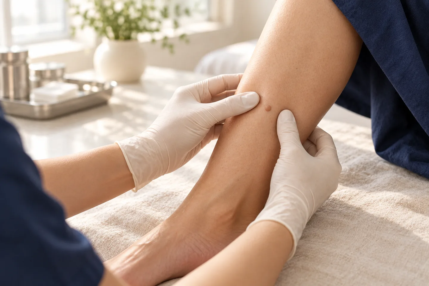
For patients considering Dermatofibroma removal in Bangkok, the first step should be confirming the diagnosis. Most typical lesions can be recognized clinically and with Dermoscopy, but a Skin Biopsy or Diagnostic Excision may be appropriate if the lesion is growing, ulcerated, unusually colored, asymmetric, or otherwise atypical.
A practical patient pathway is:
Skin lesion assessment → Dermoscopy when appropriate → Skin Biopsy if the diagnosis is uncertain → observation or Surgical Excision → Histopathology when tissue is removed
What Is a Dermatofibroma?
A Dermatofibroma, also called a Cutaneous Fibrous Histiocytoma, is a benign skin tumor that develops within the Dermis.
It is composed largely of fibroblastic and histiocytic cells together with collagen. Histologically, Dermatofibroma typically shows spindle-shaped cells and increased collagen within the Dermis.
Dermatofibromas are common in adults and may occur in people of any ethnicity. They are especially common on the legs, although they can develop elsewhere on the body.
What Does a Dermatofibroma Look Like?
A typical Dermatofibroma is:
- firm to touch;
- usually small;
- round or oval;
- brown, tan, pink, reddish, or skin-colored;
- located within the skin rather than simply sitting on the surface;
- usually slow-growing.
The lower legs are a classic location, but Dermatofibromas can also occur on the arms, shoulders, trunk, or other areas.
Some lesions are flat or only slightly raised, while others form a more obvious nodule.
The Dimple Sign
One clinical feature commonly associated with Dermatofibroma is the Dimple Sign, sometimes called the Fitzpatrick Sign.
When the skin on both sides of the lesion is gently pinched, the center may pull inward or form a small depression.
This occurs because the lesion is attached within the Dermis.
However, the Dimple Sign is not sufficient by itself to confirm the diagnosis. Atypical lesions still require proper evaluation.
What Causes Dermatofibroma?
The exact cause is not completely understood.
Dermatofibroma may represent a benign proliferation of fibrous tissue or possibly a reactive process following minor skin injury.
Some patients remember a previous:
- insect bite;
- minor trauma;
- folliculitis;
- small wound.
However, many patients have no clear preceding injury.
Dermatofibroma is not contagious and is not caused by poor hygiene.
Is Dermatofibroma Cancer?
No.
A typical Dermatofibroma is a benign skin tumor and does not normally become skin cancer.
The more important issue is that several other skin tumors can sometimes resemble Dermatofibroma.
These include:
- Dermatofibrosarcoma Protuberans or DFSP;
- Desmoplastic Melanoma;
- some melanocytic lesions;
- other spindle-cell tumors;
- occasionally basal cell carcinoma or other skin tumors.
Therefore, an atypical or changing lesion should not simply be assumed to be Dermatofibroma.
Dermatofibroma vs Dermatofibrosarcoma Protuberans
This distinction is particularly important.
Dermatofibroma is common and benign.
Dermatofibrosarcoma Protuberans or DFSP is a rare locally aggressive skin tumor that can initially resemble a plaque or nodule.
Features that may raise concern for a diagnosis other than an ordinary Dermatofibroma include:
- progressive enlargement;
- a lesion larger than expected;
- multiple nodules developing within a plaque;
- fixation or deeper extension;
- unusual clinical appearance;
- recurrence after removal.
When the diagnosis is uncertain, Skin Biopsy and Histopathology are more important than cosmetic destruction of the lesion.
Histologically, Dermatofibroma and DFSP can sometimes require Immunohistochemistry, including markers such as CD34 and Factor XIIIa, to help distinguish them.
Dermatofibroma vs Melanoma
Pigmented Dermatofibroma can sometimes appear dark brown or black and may resemble a Melanocytic Nevus or melanoma.
Some Dermatofibroma variants can be heavily pigmented and mimic melanoma.
A lesion should be evaluated if it:
- changes in shape;
- develops irregular pigmentation;
- enlarges;
- becomes asymmetric;
- ulcerates;
- bleeds without obvious trauma;
- looks different from your other skin lesions.
Dermoscopy may help determine whether the lesion has typical Dermatofibroma features or requires Biopsy.
What Does Dermatofibroma Look Like Under Dermoscopy?
Dermoscopy allows a dermatologist to examine structures beneath the surface of a skin lesion.
A classic Dermatofibroma may show:
- a central white or pale scar-like area;
- a delicate peripheral pigment network.
However, Dermoscopic patterns can vary, particularly in Skin of Colour.
Therefore, Dermoscopy supports the diagnosis but should not override concerning clinical features.
How Is Dermatofibroma Diagnosed?
Diagnosis usually begins with a clinical skin examination.
The dermatologist assesses:
- size;
- firmness;
- color;
- shape;
- location;
- depth;
- growth history;
- symptoms;
- whether the lesion shows a Dimple Sign;
- Dermoscopic structures.
Most classic Dermatofibromas can be diagnosed clinically.
When Is a Skin Biopsy Needed?
A Skin Biopsy or Diagnostic Excision may be recommended if the lesion has atypical features.
These may include:
- recent enlargement;
- ulceration;
- unusual or asymmetric pigmentation;
- asymmetric Dermoscopic structures;
- rapid change;
- an unusually large lesion;
- uncertain diagnosis.
Biopsy or Diagnostic Excision is specifically recommended when atypical features such as recent enlargement, ulceration, or asymmetric structures and colors are present.
What Does Histopathology Show?
On Histopathology, Dermatofibroma typically shows a proliferation of spindle-shaped fibrohistiocytic cells within the Dermis together with increased collagen.
Other features may include:
- collagen entrapment at the lesion edge;
- epidermal thickening;
- increased basal pigmentation;
- a storiform or whorled pattern.
There are several histological variants, including Cellular, Atypical, Epithelioid, Aneurysmal, Lipidized, and other forms.
Some variants behave differently from ordinary Dermatofibroma and may require more careful excision or follow-up.
Does Every Dermatofibroma Need Treatment?
No.
Most Dermatofibromas are harmless and can simply be observed.
Treatment may be considered when the lesion:
- is painful;
- is persistently itchy;
- is repeatedly irritated;
- catches during shaving;
- is frequently traumatized;
- causes cosmetic concern;
- has an uncertain diagnosis.
For a typical asymptomatic Dermatofibroma, reassurance is often sufficient.
Can Dermatofibroma Be Removed?
Yes.
The most definitive removal method is generally Surgical Excision, because Dermatofibroma extends into the Dermis rather than sitting only on the skin surface.
The lesion is surgically removed together with an appropriate amount of surrounding tissue, depending on its clinical characteristics.
The specimen can then be examined with Histopathology.
Surgical Excision for Dermatofibroma
Surgical Excision is usually performed under Local Anesthesia for an uncomplicated superficial lesion.
Typical steps include:
1. Clinical assessment
The dermatologist confirms whether Excision is appropriate and whether the diagnosis is sufficiently clear.
2. Local Anesthesia
Local Anesthetic is injected around the lesion.
3. Excision
The Dermatofibroma is removed through the full thickness required to remove the Dermal component.
4. Wound closure
The wound is usually closed with Suture.
5. Histopathology
The removed tissue may be sent for Histopathology to confirm the diagnosis, particularly when Diagnostic Excision is part of the reason for surgery.
Why Does Dermatofibroma Removal Usually Leave a Scar?
This is one of the most important points for patients considering cosmetic removal.
Dermatofibroma is located within the Dermis.
To remove it completely, the procedure generally needs to go deeper than a superficial skin removal.
This means that replacing the Dermatofibroma with a surgical scar may not always provide a better cosmetic result.
For a small, stable lesion that causes no symptoms, observation may be preferable to surgery.
Can Dermatofibroma Be Shaved Off?
A superficial Shave Removal may flatten the visible part of a lesion, but it may not remove the deeper Dermal component.
For this reason, superficial Shave Removal may have:
- incomplete removal;
- persistence;
- recurrence;
- scar or pigment change.
If definitive removal is the goal, Surgical Excision is generally more appropriate than simply removing the superficial portion.
Can Dermatofibroma Be Treated With Laser?
Laser may sometimes reduce pigmentation or surface prominence, but it does not necessarily remove the entire Dermal lesion.
Therefore, Laser should not be considered equivalent to complete Surgical Excision.
It is also not appropriate to destroy an undiagnosed atypical lesion simply because it resembles Dermatofibroma.
If the diagnosis is uncertain, tissue diagnosis should take priority.
Can Dermatofibroma Be Treated With Cryotherapy?
Cryotherapy is not usually a definitive method for removing a typical Dermatofibroma because the lesion extends into the Dermis.
It may alter the surface appearance but can leave part of the lesion behind.
Possible disadvantages include:
- incomplete treatment;
- recurrence;
- Hypopigmentation;
- Hyperpigmentation;
- scarring.
These pigment changes are especially relevant in darker skin tones.
Can Steroid Injection Flatten a Dermatofibroma?
Intralesional Corticosteroid has occasionally been used to reduce the prominence of selected lesions, but it does not reliably remove the Dermatofibroma.
Potential risks include:
- Skin Atrophy;
- Hypopigmentation;
- visible blood vessels;
- incomplete improvement.
It should not be presented as a guaranteed removal method.
Which Treatment Is Best?
The best management depends on the patient’s goal.
| Situation | Possible approach |
|---|---|
| Typical, asymptomatic lesion | Observation |
| Painful or repeatedly irritated lesion | Consider Surgical Excision |
| Cosmetic concern | Discuss scar versus leaving the lesion |
| Atypical or changing lesion | Skin Biopsy or Diagnostic Excision |
| Uncertain diagnosis | Histopathology rather than cosmetic destruction |
| Recurrent lesion after previous treatment | Reassessment and possible Excision |
The key question is not simply “Can it be removed?” but “Will removal leave a result that is preferable to the original lesion?”
Can Dermatofibroma Come Back After Removal?
Yes.
Recurrence can occur, particularly if the lesion is not completely removed.
Some histological variants, especially Cellular Dermatofibroma, have a higher recurrence risk than ordinary Dermatofibroma.
For this reason, an unexpectedly recurrent lesion may warrant Histopathology review or further evaluation rather than repeated superficial treatment.
What Is Cellular Dermatofibroma?
Cellular Dermatofibroma is a histological variant with greater cellularity than the common form.
It can extend more deeply and has a greater tendency to recur after Excision.
Rare aggressive behavior has also been reported, so Cellular Dermatofibroma may require more careful management and follow-up than a conventional Dermatofibroma.
Patients do not generally know whether they have this variant until Histopathology has been performed.
Are There Other Dermatofibroma Variants?
Yes.
Histological variants include:
- Cellular Dermatofibroma;
- Atypical Dermatofibroma;
- Epithelioid Dermatofibroma;
- Aneurysmal Dermatofibroma;
- Hemosiderotic Dermatofibroma;
- Lipidized Dermatofibroma;
- Clear Cell Dermatofibroma;
- Deep Fibrous Histiocytoma.
These variants are primarily Histopathologic classifications.
Some can mimic melanoma, Sarcoma, or other tumors and may require Immunohistochemistry for diagnosis.
Can You Have Multiple Dermatofibromas?
Yes.
Some people develop more than one Dermatofibroma.
Occasionally, numerous lesions can appear over a relatively short period; multiple eruptive Dermatofibromas can be associated with Immunosuppression.
A patient who suddenly develops many new Dermatofibromas should therefore discuss this with a doctor rather than assuming it is simply a cosmetic problem.
Dermatofibroma on the Leg
The lower leg is the classic location for Dermatofibroma.
A patient may notice a firm brown or pink bump that has remained relatively stable for years.
Because the lower leg can sometimes heal more slowly than areas such as the face, cosmetic removal should take scar formation and wound healing into account.
Dermatofibroma on the Face
Dermatofibroma can occur on the face, although it is less typical than on the limbs.
A facial lesion that resembles Dermatofibroma deserves careful assessment because the Differential Diagnosis may include other benign and malignant skin tumors.
If removal is considered, scar placement and cosmetic outcome should be discussed beforehand.
Dermatofibroma on the Arm or Shoulder
Dermatofibromas can also occur on the arms and shoulders.
These areas may be subject to friction, shaving, sports, or clothing, making an otherwise benign lesion bothersome.
If Surgical Excision is planned, postoperative activity may need to be modified to reduce tension on the wound.
Dermatofibroma in Asian and Darker Skin
Dermatofibroma can occur in all skin tones.
In Skin of Colour, both the clinical and Dermoscopic appearance may differ from classic textbook examples, and different Dermoscopic patterns may be seen in darker skin.
After treatment, patients with darker skin may also be more concerned about:
- Post-Inflammatory Hyperpigmentation or PIH;
- Hypopigmentation;
- prolonged redness;
- Hypertrophic Scar;
- Keloid in susceptible individuals.
These possibilities should be discussed before elective cosmetic removal.
Does Dermatofibroma Removal Hurt?
Surgical Excision is typically performed with Local Anesthesia for suitable small lesions.
The injection may sting briefly.
Once the area is numb, the procedure should not produce sharp pain, although pressure or movement may still be felt.
Some soreness is expected after the Local Anesthetic wears off.
Does Dermatofibroma Removal Need Stitches?
Complete Surgical Excision usually creates a wound that needs to be closed with Suture.
Whether the wound requires one or several layers of Suture depends on:
- lesion size;
- depth;
- body location;
- skin tension;
- wound design.
Patients should expect a linear scar rather than assuming the lesion can always be removed without stitches.
Recovery After Dermatofibroma Removal
Recovery depends on the size and location of the Excision.
Typical short-term effects may include:
- soreness;
- swelling;
- mild bruising;
- tightness around the Suture;
- temporary redness.
The wound should be protected while healing.
Wound Care After Surgical Excision
General postoperative care may include:
- keeping the wound clean;
- following Dressing instructions;
- avoiding unnecessary rubbing;
- not picking the wound;
- watching for bleeding or infection;
- avoiding excessive tension across the incision;
- returning for Suture removal or wound review when required.
Specific instructions should be based on the location and type of closure.
Can I Exercise After Dermatofibroma Removal?
Exercise restrictions depend on the surgical site.
Activities that stretch or place tension on the wound may increase the risk of:
- bleeding;
- wound separation;
- widened scar.
For example, an Excision on the shoulder, thigh, or lower leg may require temporary modification of certain activities.
Can I Swim After Dermatofibroma Removal?
Swimming is generally avoided until the wound has healed sufficiently.
This includes:
- swimming pools;
- sea water;
- hot tubs;
- natural bodies of water.
This is particularly relevant for travelers planning Dermatofibroma removal in Bangkok before continuing to beach destinations in Thailand.
Can I Fly After Dermatofibroma Removal?
Minor skin surgery does not automatically prevent air travel.
However, international patients should consider:
- wound care;
- Suture removal;
- Histopathology results;
- what to do if infection or bleeding develops;
- whether follow-up is needed.
Travel dates should ideally be discussed before surgery.
Possible Side Effects and Complications
Possible complications include:
- bleeding;
- bruising;
- infection;
- pain;
- swelling;
- wound separation;
- delayed healing;
- scar widening;
- Hyperpigmentation;
- Hypopigmentation;
- Hypertrophic Scar;
- Keloid;
- numbness;
- incomplete removal;
- recurrence.
No Dermatofibroma removal technique can guarantee a scar-free result.
When Should You See a Dermatologist?
Arrange medical evaluation if a presumed Dermatofibroma:
- grows noticeably;
- changes rapidly;
- ulcerates;
- bleeds without repeated trauma;
- develops irregular color;
- becomes asymmetric;
- is unusually large;
- becomes fixed or deeper;
- repeatedly returns after treatment;
- looks different from your other skin lesions.
These features do not automatically mean cancer, but they make diagnostic assessment more important.
What to Expect During a Dermatology Consultation in Bangkok
A consultation for a suspected Dermatofibroma should focus on diagnosis before treatment.
The dermatologist may assess:
1. How long the lesion has been present
A stable lesion present for years is different from a newly enlarging nodule.
2. Symptoms
Pain, tenderness, itching, bleeding, or rapid change should be discussed.
3. Clinical appearance
The lesion is evaluated for size, firmness, color, symmetry, and depth.
4. Dermoscopy
Dermoscopy may support a clinical diagnosis of Dermatofibroma.
5. Need for Biopsy
If features are atypical, Skin Biopsy or Diagnostic Excision may be recommended.
6. Whether removal is worthwhile
For a benign lesion, the cosmetic result of the scar should be weighed against the appearance of the original Dermatofibroma.
Dermatofibroma Removal for Tourists and International Patients in Bangkok
International patients considering removal during a short stay should plan beyond the day of the procedure.
Important considerations include:
Consultation → diagnosis → Excision if appropriate → Histopathology → wound care → Suture removal or follow-up
Before surgery, it is useful to discuss:
- planned departure date;
- whether Suture removal will occur in Thailand or overseas;
- how Histopathology results will be communicated;
- plans for swimming or strenuous activity;
- how postoperative problems would be managed after departure.
If the lesion is diagnostically uncertain, obtaining the correct diagnosis should take priority over same-day cosmetic removal.
Frequently asked questions
Is Dermatofibroma dangerous?
A typical Dermatofibroma is benign and usually harmless.
Is Dermatofibroma cancer?
No. Ordinary Dermatofibroma is not skin cancer.
Can Dermatofibroma turn into cancer?
Typical Dermatofibroma does not normally transform into cancer. The more important concern is distinguishing it from other tumors that may look similar.
Does Dermatofibroma need treatment?
Usually not. Observation is often appropriate when the diagnosis is clear and the lesion causes no symptoms.
Why is Dermatofibroma hard?
It contains fibrous tissue and collagen within the Dermis, which gives it its characteristic firm feel.
What is the Dimple Sign?
It is an inward depression that may appear when the skin around a Dermatofibroma is pinched.
Can Dermatofibroma itch?
Yes. Some lesions can be itchy, painful, or tender.
Can Dermatofibroma grow?
It may grow slowly, but significant recent enlargement should be assessed.
What is the best way to remove Dermatofibroma?
Complete Surgical Excision is generally the most definitive removal method.
Can Dermatofibroma be removed with Laser?
Laser may alter the lesion's appearance but does not necessarily remove the full Dermal component.
Can Dermatofibroma be frozen with Cryotherapy?
Cryotherapy is generally not considered a definitive way to remove the entire lesion and may cause pigment change.
Can Dermatofibroma be removed with Shave Removal?
Superficial Shave Removal may leave deeper Dermatofibroma tissue behind.
Does Dermatofibroma removal leave a scar?
Yes. Complete removal usually requires Surgical Excision and therefore leaves a surgical scar.
Can Dermatofibroma return after surgery?
Yes, particularly if it is incompletely removed or if it is a histological variant with a higher recurrence tendency.
What is Cellular Dermatofibroma?
It is a histological variant that has a greater tendency to recur and may require more careful management.
Is Dermatofibroma the same as DFSP?
No. Dermatofibroma is benign, while Dermatofibrosarcoma Protuberans is a different locally aggressive tumor.
Can Dermoscopy diagnose Dermatofibroma?
Dermoscopy can strongly support the diagnosis, but Skin Biopsy may still be required for atypical lesions.
When does Dermatofibroma need a Biopsy?
Biopsy may be appropriate for lesions showing recent growth, ulceration, atypical color, asymmetry, or uncertain diagnosis.
Should an excised Dermatofibroma be sent for Histopathology?
When removal is performed for diagnostic uncertainty or atypical features, Histopathology is important for confirming the diagnosis.
Can Dermatofibroma occur on the face?
Yes, although the lower legs are much more typical.
Can Dermatofibroma occur in darker skin?
Yes. It occurs across ethnicities, and Dermoscopic appearances may differ in Skin of Colour.
Can you have multiple Dermatofibromas?
Yes. Multiple lesions can occur, and sudden development of many lesions has been associated with Immunosuppression.
Can I exercise after Dermatofibroma removal?
It depends on the location and wound size. Activities that place tension on the incision may need to be limited temporarily.
Can I swim after Dermatofibroma removal?
Usually not until the wound has healed sufficiently.
Can tourists have Dermatofibroma removal in Bangkok?
Potentially yes, but timing of Suture removal, Histopathology results, and wound care should be planned before traveling onward.
Related conditions
-
Mole Removal Mole Removal Many growths that patients call “moles” are benign and can be removed because they catch on clothing, become repeatedly irritated, or are cosmetically unwanted. Others may actually be Seborrheic Keratosis, Skin Tag, Wart, Actinic Keratosis, or another skin condition. Occasionally, a lesion that looks like an ordinary mole or growth can represent Melanoma, Basal Cell Carcinoma, SCC, or another skin cancer.

-
Dermoscopy Dermoscopy Dermoscopy is a non-invasive examination technique that allows a dermatologist to inspect skin lesions with greater detail than is possible with the naked eye alone.

-
Skin Biopsy (การวินิจฉัย / Diagnosis) Skin Biopsy (การวินิจฉัย / Diagnosis) A skin biopsy is a minor medical procedure in which a dermatologist removes a small sample of skin so it can be examined under a microscope. It is one of the most important diagnostic tools in dermatology because many skin conditions cannot be confirmed by appearance alone.

-
Excisional Biopsy Excisional Biopsy An excisional biopsy means completely cutting out a skin lesion, usually through the full thickness of the skin. The removed tissue is then examined under a microscope.
