
































Excisional Biopsy
An excisional biopsy means completely cutting out a skin lesion, usually through the full thickness of the skin. The removed tissue is then examined under a microscope.
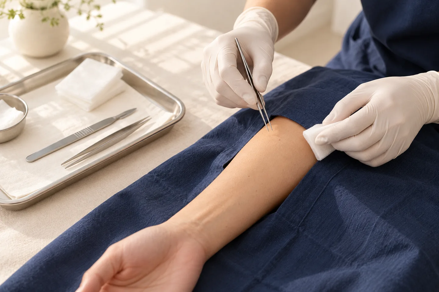
Smaller lesions are commonly removed using an elliptical incision and the wound is closed with stitches. Larger defects may occasionally require more complex reconstruction such as a skin flap or graft.
An excisional biopsy can have two roles:
Diagnostic: It provides tissue so the pathologist can determine exactly what the lesion is.
Potentially therapeutic: In some cases, the procedure may remove the entire abnormal lesion.
However, these two concepts should not be confused. A lesion being visibly removed does not automatically mean treatment is complete. If pathology shows skin cancer, further surgery or another treatment may still be required depending on the diagnosis, margin status and other pathological features.
Why Would a Dermatologist Recommend an Excisional Biopsy?
Many skin lesions can be diagnosed clinically and do not need surgery.
A dermatologist may consider excisional biopsy when:
- A mole or pigmented lesion has suspicious features.
- Melanoma is included in the differential diagnosis.
- Examination of the entire lesion is important.
- A small tumor can reasonably be removed completely.
- A lesion is growing, changing or repeatedly bleeding.
- Previous assessment has not established a secure diagnosis.
- The relationship between a tumor and its margins is important.
- A symptomatic lesion requires removal but should also be examined histologically.
The biopsy method should be selected according to the suspected diagnosis rather than simply choosing the smallest or easiest procedure. Biopsy type and sampling site can substantially influence the usefulness of the pathology specimen.
What Skin Lesions May Require an Excisional Biopsy?
Suspicious Moles and Pigmented Lesions
A changing mole is one of the most common reasons patients search for information about excisional biopsy.
Features that deserve dermatological assessment include:
- A new or unusual pigmented lesion
- Progressive enlargement
- Changing shape
- Increasing asymmetry
- Irregular borders
- Multiple or changing colors
- Unexplained bleeding
- Ulceration
- A lesion that appears noticeably different from the patient’s other moles
These findings do not automatically mean melanoma. Benign moles can also be irregular or change gradually.
The purpose of evaluation is to identify which lesions need biopsy and which can reasonably be monitored.
Excisional Biopsy for Suspected Melanoma
Biopsy technique is especially important when melanoma is suspected.
An excisional biopsy, in which the whole abnormal area is removed with a small margin of normal skin, is usually the preferred biopsy method for suspected melanoma. The reason is that the pathologist needs adequate tissue to evaluate the lesion accurately, including how deeply a melanoma has invaded if one is present.
An incisional or other partial biopsy may still be appropriate when complete initial removal is difficult—for example, because the lesion is very large or situated in an anatomically challenging area.
Excisional biopsy is not the same as definitive melanoma surgery
This distinction is essential.
The initial procedure is primarily performed to establish the diagnosis and obtain the pathological information needed for treatment planning.
If melanoma is confirmed, the patient generally enters a separate treatment pathway. Surgery remains central to melanoma management, and further excision may be required according to the pathological findings and stage.
Basal Cell Carcinoma
Basal cell carcinoma is a form of skin cancer that can present in several different ways.
A lesion suspected of basal cell carcinoma does not always require an excisional biopsy as the initial test. A punch or shave biopsy may sometimes provide enough tissue to establish the diagnosis or determine the histological growth pattern.
In selected small lesions, complete surgical removal may be reasonable.
If pathology confirms basal cell carcinoma, further management depends on factors such as:
- Histological subtype
- Location
- Size
- Clinical borders
- Margin status
- Whether the lesion is recurrent
The initial biopsy therefore needs to be interpreted together with the clinical lesion.
Squamous Cell Carcinoma
A suspicious squamous cell carcinoma may also be assessed by punch, shave, incisional or excisional biopsy depending on the lesion.
The dermatologist needs enough tissue to answer the relevant diagnostic question, including whether invasive disease is present and, in some cases, its depth or growth pattern.
Punch biopsy is useful for many suspected basal cell and squamous cell carcinomas because it provides a deeper specimen, although it generally should not be viewed as a method for completely removing a skin cancer.
Benign Skin Lesions
Excisional biopsy is not limited to cancer.
Selected benign lesions may also be completely removed for diagnosis or because they are:
- Painful
- Repeatedly irritated
- Frequently inflamed
- Growing
- Difficult to diagnose with confidence
- Bothersome to the patient
Examples can include selected moles, cysts and benign skin tumors.
However, benign lesions do not automatically need surgical removal. If a lesion can confidently be diagnosed as harmless and is not causing a problem, observation may be reasonable.
Excisional Biopsy vs Punch Biopsy
These procedures answer different clinical questions.
| Excisional Biopsy | Punch Biopsy | |
|---|---|---|
| Tissue removed | Entire visible lesion | Small cylindrical sample |
| Depth | Usually full-thickness | Usually includes epidermis and dermis and may reach superficial subcutaneous tissue |
| Main purpose | Evaluate the entire lesion | Obtain a representative sample |
| Stitches | Usually required | Sometimes required |
| Useful for | Selected suspicious moles and small tumors | Inflammatory diseases and selected tumors |
| Main limitation | Larger wound and scar | Sampling may not represent the whole lesion |
A punch biopsy can be very useful when the dermatologist needs to see changes occurring through several layers of the skin. This is particularly relevant for many inflammatory skin diseases and selected tumors.
For a suspicious melanocytic lesion, however, sampling only a small portion can miss important pathology. Complete excision is generally preferred when melanoma is a concern and removal of the whole lesion is practical.
Excisional Biopsy vs Incisional Biopsy
An excisional biopsy removes the entire lesion.
An incisional biopsy removes only part of the lesion.
Incisional biopsy may be preferable when:
- The lesion is too large to remove completely as an initial procedure.
- Complete removal would create an unnecessarily large defect.
- The location makes complete initial excision difficult.
- A representative sample is sufficient to establish the diagnosis.
For suspected melanoma, incisional biopsy can be used when the entire lesion cannot easily be removed, but complete excision is generally preferred when feasible.Excisional Biopsy vs Shave Biopsy
A shave biopsy removes tissue horizontally from the skin surface.
The depth can vary considerably. A superficial shave removes mainly the upper layers, while a deeper saucerization biopsy obtains substantially more tissue.
Shave biopsy can be appropriate for many superficial or raised lesions. However, a superficial biopsy has limitations when the pathologist needs to determine whether abnormal cells extend deeper into the skin.
The appropriate choice should depend on the suspected diagnosis—not simply which procedure leaves the smallest wound.
How Does a Dermatologist Decide Which Biopsy Is Best?
The dermatologist may consider:
- Suspected diagnosis
- Whether the lesion is pigmented
- Possibility of melanoma or another skin cancer
- Size
- Depth
- Anatomical location
- Whether the entire lesion must be assessed
- Whether only a representative sample is required
- Risk of scarring
- Important structures beneath or beside the lesion
- Whether special laboratory testing may be required
The principle is straightforward:
The biopsy should obtain enough appropriate tissue to answer the diagnostic question while avoiding unnecessary surgery.
The smallest biopsy is not necessarily the best biopsy.
What Happens Before an Excisional Biopsy?
Clinical Examination
The dermatologist first examines the lesion and surrounding skin.
History may include:
- How long the lesion has been present
- Whether it has changed
- Bleeding or ulceration
- Pain or itching
- Previous skin cancer
- Previous biopsies
- Current medication
- Blood-thinning medication
- Drug or local-anesthetic allergy
- Previous problems with wound healing
- Previous hypertrophic scars or keloids
It is recommended to inform the clinician about medications that can affect bleeding and relevant medical conditions before skin excision.
Dermoscopy
For many pigmented lesions and selected non-pigmented tumors, the dermatologist may examine the lesion with a dermoscope before deciding whether biopsy is required.
Dermoscopy allows structures beneath the skin surface to be assessed in greater detail, but it does not replace histopathology when tissue diagnosis is necessary.
How Is an Excisional Biopsy Performed?
1. Marking the Lesion
The dermatologist identifies the lesion and plans the incision.
Small lesions are often removed through an elliptical or oval-shaped excision so the resulting defect can be closed as a line.
2. Cleaning and Local Anesthesia
The skin is cleaned and local anesthetic is injected.
The injection may briefly sting or burn. Once the anesthetic has taken effect, patients should not normally feel sharp cutting pain during the procedure.
Routine excisional skin biopsies can generally be carried out while the patient is awake under local anesthesia.
3. Removing the Lesion
A scalpel is used to cut through the full thickness of skin and remove the lesion.
Depending on the reason for surgery, the specimen may include a small amount of surrounding clinically normal skin.
For suspected melanoma, the purpose of the initial diagnostic excision is to obtain adequate tissue for diagnosis. This should not be confused with the larger definitive surgical margin that may subsequently be required if melanoma is confirmed.
4. Controlling Bleeding
Bleeding is controlled using appropriate surgical techniques, which may include pressure or electrosurgical methods.
5. Closing the Wound
Most excisional biopsies are closed with sutures.
Closure may involve:
- Surface stitches
- Absorbable stitches underneath the skin
- A combination of both
For larger defects, repair with a skin flap or graft may occasionally be required.6. Sending the Tissue for Histopathology
The removed specimen is sent to a pathology laboratory.
For routine examination, tissue is generally preserved in formalin, processed into thin sections, stained and examined microscopically by a pathologist or dermatopathologist.
This step is what transforms simple lesion removal into a diagnostic biopsy.
What Is Histopathology?
Histopathology is the microscopic examination of tissue.
The pathologist evaluates the architecture of the lesion and the appearance of individual cells.
Depending on the diagnosis, the report may provide information about:
- The exact diagnosis
- Whether a lesion is benign or malignant
- Tumor subtype
- Tumor depth or thickness when relevant
- Other pathological risk factors
- Whether abnormal tissue reaches the edges of the specimen
The clinical diagnosis supplied to the laboratory also matters. Pathology interpretation benefits from information such as lesion site, duration, suspected diagnosis and relevant history.
What Do “Clear Margins” Mean?
The margin is the edge of the tissue removed during surgery.
A pathology report may describe the examined margins as clear or involved.
A clear or negative margin generally means abnormal cells are not seen extending to the examined specimen edge.
An involved or positive margin means the abnormal process reaches an examined edge of the specimen.
However, margin interpretation depends on:
- The diagnosis
- Biopsy technique
- Specimen orientation
- How the tissue is processed
- Clinical appearance of the original lesion
Dermatopathology can help determine whether excision margins are adequate, but the pathology report still needs to be interpreted alongside the patient’s clinical findings.
If the Excisional Biopsy Removes Everything, Is Treatment Finished?
Sometimes—but not always.
For a benign lesion, complete excision may require no further treatment.
For malignant lesions, however, the amount removed during a diagnostic biopsy may not meet the margin or treatment requirements for definitive cancer surgery.
This is particularly important for melanoma.
Therefore:
“The lesion has been removed” does not necessarily mean “the treatment has been completed.
”
The next step should be based on the pathology diagnosis.
What Happens If Pathology Shows Basal Cell Carcinoma?
If basal cell carcinoma is diagnosed, management depends on factors including:
- Histological subtype
- Location
- Size
- Margin status
- Clinical tumor borders
- Previous treatment
- Whether the lesion is recurrent
Further excision or another treatment may be recommended depending on the overall situation.
What Happens If Pathology Shows Squamous Cell Carcinoma?
Further treatment may also be necessary after a diagnosis of squamous cell carcinoma.
Treatment planning can be influenced by:
- Tumor size
- Anatomical location
- Depth
- Histological differentiation
- Margin status
- Other pathological risk features
- Immune status of the patient
The pathology report should therefore be considered part of the treatment-planning process rather than simply a confirmation of the diagnosis.
What Happens If the Excisional Biopsy Shows Melanoma?
Melanoma requires a separate treatment and staging pathway.
The pathology report may contain information such as:
- Melanoma in situ versus invasive melanoma
- Breslow thickness
- Ulceration
- Margin status
- Other pathological characteristics
Surgery remains the cornerstone of treatment for cutaneous melanoma. Further surgery is generally planned according to the pathological findings, and staging procedures may be considered in appropriate patients.
This is why an appropriate initial biopsy is so important: the pathology from that specimen can influence what happens next.
Does an Excisional Biopsy Leave a Scar?
Yes.
It is not possible to completely cut a lesion out of the full thickness of skin without producing some degree of permanent scar.
A well-planned procedure aims to make the scar as acceptable as reasonably possible, but “scar-free excision” is not a realistic promise.
Scar appearance depends on:
- Size of the lesion and excision
- Location
- Skin tension
- Direction of the incision
- Individual healing tendency
- Wound infection or separation
- Repeated stretching of the wound
- Tendency to develop hypertrophic or keloid scars
Some body areas naturally develop more noticeable scars than others.
What Will the Scar Look Like?
An elliptical excision that is closed directly usually produces a linear scar rather than a round defect.
Early scars may appear:
- Pink or red
- Slightly raised
- Firm
- More noticeable than expected
The appearance typically evolves as scar remodeling occurs.
Some patients develop a wider scar or a thicker hypertrophic or keloid scar despite appropriate surgical technique. People with an abnormal scarring response can develop larger or thicker scars following excision.
Recovery After Excisional Biopsy
Recovery varies substantially according to the size and location of the wound.
A small biopsy on a relatively immobile area is different from an excision over the shoulder, back, knee or another area that is repeatedly stretched.
Patients may experience:
- Mild tenderness
- Minor swelling
- Bruising
- Tightness from stitches
The surgical wound initially heals over a relatively short period, while the scar continues changing for much longer.
Will I Need Stitches?
Usually.
Because an excisional biopsy removes the entire lesion through the full thickness of skin, the wound is commonly closed with sutures.
The number and type of stitches depend on:
- Length of the wound
- Depth
- Location
- Skin tension
- Surgical technique
Some sutures beneath the skin dissolve naturally, while surface stitches may need to be removed.
How Long Do Stitches Stay In?
There is no single removal time that applies to every biopsy.
Timing depends on:
- Face versus trunk or limbs
- Wound tension
- Healing
- Type of suture
- Individual factors
Patients should follow the date provided by the treating clinician rather than using a general internet timetable.
For tourists or patients leaving Bangkok soon after surgery, the stitch-removal plan should ideally be arranged before the procedure.
Can I Shower After an Excisional Biopsy?
Wound-care instructions differ according to the dressing, surgical site and closure technique.
Patients should follow the specific instructions provided after surgery regarding:
- When the dressing can be removed
- When the wound can get wet
- How to clean it
- Whether an ointment should be applied
- Whether a replacement dressing is required
Avoid applying unrecommended antiseptics, herbal preparations or cosmetic products directly to a fresh surgical wound.
Can I Exercise After an Excisional Biopsy?
Exercise may pull on a fresh wound, particularly when the incision is located on:
- Shoulder
- Upper back
- Chest
- Abdomen
- Arms or legs
- Near a joint
Excessive tension can contribute to bleeding, wound separation or a wider scar.
Activity restrictions should therefore be based on the wound location rather than a single universal rule.
Ask your dermatologist specifically about gym exercise, running, swimming and sports if these are part of your usual routine.
Possible Side Effects and Complications
Excisional biopsy is generally a minor surgical procedure, but complications can occur.
Pain
Tenderness may develop after the local anesthetic wears off.
Bleeding
Minor bleeding or spotting can occur.
Patients taking anticoagulant, antiplatelet or other medicines that affect bleeding should tell the clinician before surgery. Do not stop prescription medication independently just because a biopsy is planned.
Bruising or Hematoma
Blood can occasionally accumulate beneath the wound.
Infection
Possible warning signs include:
- Increasing redness
- Increasing warmth
- Progressive swelling
- Increasing pain
- Pus
- Fever or feeling generally unwell
Wound Separation
The edges of the wound can separate, particularly if the area is under significant tension or if infection interferes with healing.
Permanent Scar
Some scar is expected following full-thickness excision.
Hypertrophic or Keloid Scar
Some patients are predisposed to developing thick raised scars.
Altered Sensation
Cutting through the skin can affect very small sensory nerves. Temporary numbness, tingling or altered sensation may occur around some surgical sites.
Incomplete Microscopic Removal
Even if the entire lesion appears to have been removed clinically, pathology may show that abnormal tissue extends to an examined margin.
Additional treatment can then be considered according to the diagnosis.
When Should You Contact a Doctor After the Procedure?
Seek medical advice if you develop:
- Persistent bleeding despite firm pressure
- Rapidly increasing swelling
- Increasing rather than improving pain
- Spreading redness
- Pus or foul-smelling drainage
- Fever
- Wound separation
- Significant loss of sensation or another unexpected neurological symptom
- Any rapidly worsening postoperative problem
A wound that looks increasingly inflamed several days after surgery should not simply be assumed to represent normal healing.
How Long Do Pathology Results Take?
There is no reliable universal turnaround time.
Some specimens are straightforward, while others may require:
- Additional tissue sections
- Special stains
- Immunohistochemistry
- Specialist dermatopathology review
- Additional consultation
Special stains or other investigations may sometimes be required before the diagnosis is finalized.
For this reason, patients should ask the treating clinic when the report is realistically expected.
Pathology Planning for Tourists and International Patients
This is particularly important in Bangkok, where some patients are visiting Thailand only temporarily.
Before undergoing excisional biopsy, clarify:
- When the pathology report is expected
- How the result will be communicated
- Whether an online follow-up is possible if you leave Thailand
- When stitches need to be removed
- Who can remove the stitches after you return home
- Whether you need copies of pathology reports or clinical photographs
- What happens if additional treatment is recommended
Ideally, the follow-up pathway should be planned before the tissue is removed, not after the patient has already left the country.
Can I Fly After an Excisional Biopsy?
A small skin excision does not automatically prevent air travel.
The practical issues are usually:
- Wound care
- Bleeding risk
- Activity restrictions
- Stitch removal
- Access to medical care if complications occur
- Receiving the pathology result
- Potential need for further surgery
If you have a flight soon after the planned procedure, tell the dermatologist before surgery so the timing and follow-up plan can be considered.
Can an Excisional Biopsy Miss the Diagnosis?
Removing the entire visible lesion generally reduces the sampling limitation associated with taking only a small portion of a heterogeneous lesion.
However, pathology is still not interpreted in isolation.
Accurate diagnosis depends on:
- Correct lesion being selected
- Appropriate biopsy technique
- Adequate specimen
- Correct specimen handling
- Clinical information supplied to the pathologist
- Microscopic interpretation
- Correlation between pathology and the clinical appearance
Special emphasis is placed on clinicopathological correlation and providing the pathology laboratory with the suspected diagnosis, lesion site and relevant history.
Do All Changing Moles Need to Be Removed?
No.
A dermatologist may recommend:
- Observation
- Dermoscopic monitoring
- Clinical photography
- Sequential digital monitoring
- Biopsy
- Complete excision
depending on the lesion.
Removing every mole unnecessarily results in unnecessary surgery and scars.
Conversely, repeatedly destroying or cosmetically treating a suspicious pigmented lesion without first establishing the diagnosis can be inappropriate.
Diagnosis should come before destruction of a suspicious lesion.
When Should You Have a Mole or Skin Lesion Checked?
Consider dermatological evaluation if a lesion:
- Is new and unusual
- Is changing progressively
- Is growing
- Develops irregular pigmentation
- Changes shape
- Develops irregular borders
- Repeatedly bleeds
- Becomes ulcerated
- Does not heal
- Forms a persistent crust
- Becomes a persistent enlarging lump
- Looks significantly different from your other moles
These signs are not proof of skin cancer.
They are reasons to have the lesion assessed.
What to Expect During an Excisional Biopsy Consultation in Bangkok?
A useful consultation should answer more than simply whether a lesion “can be removed.
”
The dermatologist first needs to determine what the lesion might be and what information the biopsy needs to provide.
History
You may be asked about:
- When you first noticed the lesion
- Whether it has changed
- Previous treatment
- Symptoms
- Previous skin cancers
- Family or personal melanoma history where relevant
- Medication
- Bleeding tendency
- Allergies
- Previous wound-healing problems
Examination
The lesion is assessed for:
- Size
- Shape
- Color
- Surface
- Borders
- Depth
- Anatomical location
Dermoscopy may be used when appropriate.
Choosing the Biopsy Method
Possible options can include:
- Shave biopsy
- Punch biopsy
- Incisional biopsy
- Excisional biopsy
Each has advantages and limitations.
A more extensive procedure is not automatically a better procedure.
Discussion Before Surgery
Before agreeing to excisional biopsy, patients should understand:
- Why biopsy is recommended
- Whether complete excision is planned
- Alternative biopsy techniques
- Expected wound and scar
- Whether stitches are needed
- Possible complications
- Wound care
- When pathology is expected
- What further treatment could be required
Excisional Biopsy in Bangkok: Key Takeaway
An excisional biopsy is both surgery and a diagnostic test.
The most important benefit is not simply that a visible lesion disappears. It is that the entire lesion can be examined under the microscope, giving the pathologist information that can determine whether the lesion is benign, malignant or requires further treatment.
For suspicious moles and possible melanoma, the initial biopsy technique matters because inadequate sampling can limit the pathological information needed for subsequent management. When feasible, excisional biopsy is usually preferred for a lesion suspected of melanoma.
For patients in Bangkok—particularly tourists and international patients—a complete plan should include not only the biopsy itself but also histopathology, wound care, stitch removal, communication of results and a clear pathway for further treatment if required.
Frequently asked questions
What is an excisional biopsy?
It is a surgical biopsy in which the entire visible skin lesion is removed and submitted for microscopic examination.
Is an excisional biopsy painful?
Local anesthetic is normally used. The injection can briefly sting, but sharp cutting pain should not normally be felt once the area is adequately numb.
Is excisional biopsy the same as mole removal?
Not necessarily.
“Mole removal” describes removing a mole, which can be done using different techniques and for different reasons. An excisional biopsy specifically removes the lesion in a way that allows it to be sent for histopathological diagnosis.
Does an excisional biopsy remove the whole lesion?
By definition, the intention is to remove the entire visible lesion. Microscopic examination can nevertheless show abnormal cells extending to an examined margin.
Is excisional biopsy preferred for suspected melanoma?
Usually, when removing the entire suspicious lesion is technically feasible. Excisional biopsy is usually described as the preferred biopsy method for suspected melanoma.
Can punch biopsy be used instead?
Sometimes, depending on the lesion. Punch biopsy is valuable for many inflammatory diseases and selected tumors. However, partial sampling of a suspicious melanocytic lesion can miss diagnostically important areas.
Will I need stitches?
Usually, yes. Excisional biopsies commonly create a full-thickness wound that is closed with sutures.
Will I have a scar?
Yes. Full-thickness surgical removal produces some degree of permanent scarring.
What happens to the removed tissue?
It is sent to a pathology laboratory, processed and examined microscopically.
What if the result is benign?
If the diagnosis is benign and no additional management is required, observation may be all that is needed after the wound has healed.
What if the result is skin cancer?
The next step depends on the type of cancer, pathology findings, location and clinical situation. Additional surgery or another treatment may be recommended.
If melanoma was completely removed during biopsy, is additional surgery still necessary?
Often, yes. Diagnostic biopsy and definitive melanoma treatment are separate steps. Surgery remains central to melanoma treatment and further excision is generally planned according to the pathology.
What does a positive margin mean?
It means abnormal cells extend to an examined edge of the tissue specimen. Whether additional treatment is necessary depends on the diagnosis and clinical situation.
How long does an excisional biopsy take?
The actual excision may be relatively brief, but appointment time also includes examination, marking, local anesthesia, bleeding control, wound closure and dressing. The duration varies according to complexity.
When can I exercise again?
It depends on the wound location and tension. Your dermatologist should give individualized advice.
Can international patients have an excisional biopsy in Bangkok?
Potentially, yes, but pathology reporting, wound review and stitch removal should be planned carefully if the patient is leaving Thailand soon after the procedure.
Related conditions
-
Skin Biopsy (การวินิจฉัย / Diagnosis) Skin Biopsy (การวินิจฉัย / Diagnosis) A skin biopsy is a minor medical procedure in which a dermatologist removes a small sample of skin so it can be examined under a microscope. It is one of the most important diagnostic tools in dermatology because many skin conditions cannot be confirmed by appearance alone.

-
Punch Biopsy Punch Biopsy A punch biopsy uses a small circular cutting instrument to remove a cylindrical sample of skin. Unlike a superficial shave biopsy, a punch biopsy generally provides a full-thickness specimen containing the epidermis and dermis and often extending into superficial subcutaneous tissue. This allows the pathologist to examine the architecture of different layers of the skin.

-
Shave Biopsy Shave Biopsy A shave skin biopsy is a common dermatology procedure used to remove a superficial portion of a skin lesion for examination under a microscope. It is usually performed under local anesthesia during an outpatient dermatology visit and often does not require stitches.
-
Histopathology Histopathology Histopathology is the microscopic examination of tissue to identify disease. In dermatology, this usually means examining a piece of skin removed during a skin biopsy or surgical excision.
