































Histopathology
Histopathology is the microscopic examination of tissue to identify disease. In dermatology, this usually means examining a piece of skin removed during a skin biopsy or surgical excision.
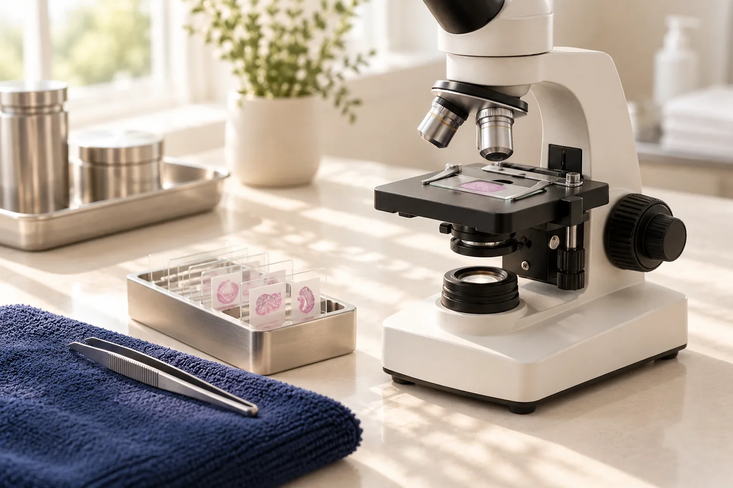
A dermatologist may suspect a diagnosis from the appearance of a lesion, its history and dermoscopy, but some conditions cannot be diagnosed reliably from appearance alone. Histopathology allows a pathologist to examine the architecture and individual cells within the tissue and can help diagnose skin cancer, inflammatory skin disease, certain infections and many other skin conditions. For suspected skin cancer, microscopic examination of biopsy tissue is required to establish the diagnosis.
For patients in Bangkok—including Thai patients, expatriates, tourists and international patients—understanding histopathology is particularly useful when a dermatologist recommends a biopsy, when a pathology result contains unfamiliar terminology, or when further treatment depends on the microscopic diagnosis.
What Is Histopathology?
Histopathology is the study of structural and cellular changes in tissue caused by disease.
The word can be understood in three parts:
Histo refers to tissue.
Pathology refers to the study of disease.
Histopathology therefore means examining diseased tissue, usually under a microscope.
In skin disease, the more specific term dermatopathology refers to the microscopic study and interpretation of skin specimens. Dermatopathology combines microscopic findings with clinical information about the patient’s skin disease.
What Is a Dermatopathologist?
A dermatopathologist is a doctor with specialized expertise in diagnosing skin disease by examining tissue under a microscope.
Depending on the healthcare system and case, a skin biopsy may be interpreted by:
- A general pathologist
- A histopathologist
- A dermatopathologist
Dermatopathology can be particularly valuable because skin diseases often share similar microscopic patterns. The microscopic appearance sometimes needs to be interpreted alongside photographs, lesion location, symptoms, duration and the dermatologist’s suspected diagnoses.
Why Is Histopathology Important in Dermatology?
Many skin disorders look similar from the outside.
For example, a red scaly patch could represent an inflammatory condition, precancerous change or skin cancer. A dark lesion could be a benign mole, seborrheic keratosis, pigmented basal cell carcinoma or melanoma.
Histopathology can provide information that cannot be seen during ordinary examination.
It may help:
- Confirm a clinical diagnosis
- Distinguish benign from malignant lesions
- Identify the type of skin cancer
- Assess how deeply a tumor has invaded
- Evaluate whether an excised lesion reaches the surgical margins
- Distinguish between similar inflammatory skin diseases
- Identify certain infectious organisms
- Guide further treatment
A pathology report can therefore influence both diagnosis and treatment planning.
Histopathology vs Skin Biopsy: What Is the Difference?
These terms are closely related but are not the same.
A skin biopsy is the procedure used to remove tissue from the patient.
Histopathology is what happens after that tissue is processed and examined microscopically.
A simple way to think about it is:
Skin examination → biopsy → laboratory processing → microscopic examination → pathology report → treatment or follow-up plan
The biopsy provides the tissue. Histopathology provides the microscopic diagnosis.
When Might a Dermatologist Recommend Histopathology?
Histopathology is commonly considered when the diagnosis cannot be established confidently from examination alone, when skin cancer is suspected, or when the result would change management.
Examples include:
- Suspicious mole
- Changing pigmented lesion
- Possible melanoma
- Possible basal cell carcinoma
- Possible squamous cell carcinoma
- Persistent ulcer or non-healing lesion
- Uncertain skin tumor
- Atypical mole
- Persistent unexplained rash
- Possible autoimmune skin disease
- Blistering disease
- Vasculitis
- Unusual inflammatory skin condition
- Suspected deeper skin disease
- Selected infections
- Scarring hair loss
- Certain nail disorders
A biopsy may also be used to determine whether a previously diagnosed tumor has been completely excised.
Is Histopathology Only Used for Skin Cancer?
No.
Skin cancer is one of the most important uses of histopathology, but it is only one part of dermatopathology.
Histopathology can also help evaluate:
- Eczema-like disorders
- Psoriasis-like eruptions
- Lichen planus
- Autoimmune connective-tissue diseases
- Vasculitis
- Blistering disorders
- Granulomatous inflammation
- Drug eruptions
- Hair and scalp disease
- Certain infections
- Unusual skin growths
Some diagnoses can be made clinically without biopsy. Others are much more difficult to distinguish without microscopic examination.
Is Histopathology Necessary for Suspected Skin Cancer?
When a lesion is suspected to be skin cancer, a skin biopsy and microscopic examination are generally required for definitive diagnosis.
Standard medical guidelines state that biopsy is the way to establish whether a suspicious lesion is skin cancer and, if cancer is present, determine the type.
This distinction is important.
A dermatologist may say:
“This looks very suspicious for basal cell carcinoma.
”
“This could be melanoma.
”
“This lesion appears benign.
”
But when cancer is genuinely suspected, the final diagnosis usually comes from tissue examination rather than visual inspection alone.
Histopathology for Melanoma
Histopathology is particularly important when melanoma is suspected.
A suspicious lesion is biopsied and examined microscopically for melanoma cells.
If melanoma is identified, the pathology report may contain clinically important information such as:
- Melanoma type
- Tumor thickness
- Ulceration
- Mitotic activity in relevant situations
- Margin involvement
- Other microscopic features important for staging and management
The depth of melanoma is particularly important because it influences subsequent treatment and staging decisions.
Histopathology for Basal Cell Carcinoma
Basal cell carcinoma can have several clinical and microscopic subtypes.
A lesion may appear:
- Pearly
- Pink
- Ulcerated
- Crusted
- Pigmented
- Scar-like
Histopathology confirms whether basal cell carcinoma is present and can provide information about the tumor pattern that may influence treatment planning.
A lesion should not simply be assumed to be basal cell carcinoma from appearance when diagnostic uncertainty remains.
Histopathology for Squamous Cell Carcinoma
Squamous cell carcinoma may appear as:
- A growing firm lesion
- A rough or thickened plaque
- A crusted growth
- A bleeding lesion
- A non-healing ulcer
Histopathology helps distinguish invasive squamous cell carcinoma from other lesions such as actinic keratosis, squamous cell carcinoma in situ and benign keratinizing growths.
Biopsy remains necessary when the diagnosis needs confirmation.
Histopathology for Suspicious Moles
A mole may look unusual clinically or under dermoscopy without necessarily being melanoma.
Possible pathology results after biopsy include:
- Benign melanocytic nevus
- Atypical or dysplastic nevus
- Melanoma in situ
- Invasive melanoma
- Another pigmented lesion
This is why a suspicious mole should ideally be assessed before it is destroyed cosmetically.
When melanoma is a meaningful possibility, obtaining tissue for pathological examination is more important than simply removing the visible pigmentation.
Can Histopathology Diagnose Inflammatory Skin Disease?
Yes, but the process can be more complicated than diagnosing some tumors.
Many inflammatory skin diseases produce overlapping microscopic patterns. Histopathology may therefore:
- Confirm a suspected diagnosis
- Narrow the differential diagnosis
- Exclude another disease
- Describe a reaction pattern rather than give one definitive diagnosis
DermNet emphasizes that inflammatory skin pathology often requires clinicopathological correlation—combining what the pathologist sees under the microscope with what the dermatologist sees on the patient.
What Is Clinicopathological Correlation?
Clinicopathological correlation means combining:
Clinical information + physical examination + photographs or dermoscopy when relevant + microscopic findings
This is one of the most important concepts in dermatopathology.
For example, under the microscope several diseases may show a similar inflammatory pattern.
The pathologist may need to know:
- Where the rash is located
- How long it has been present
- Whether it itches
- Whether it is painful
- Whether blisters are present
- Which medications the patient uses
- Whether the lesion was treated before biopsy
- What diagnoses the dermatologist is considering
The final diagnosis may become much clearer when the clinical and microscopic information are interpreted together.
What Types of Skin Biopsy Can Be Sent for Histopathology?
Different biopsy techniques provide different amounts and depths of tissue.
Punch Biopsy
A punch biopsy uses a small circular instrument to remove a cylindrical sample of skin.
It can include:
- Epidermis
- Dermis
- Sometimes subcutaneous tissue
Punch biopsy is commonly useful for inflammatory diseases and many tumors because it gives the pathologist a cross-sectional view of the skin.
Shave Biopsy
A shave biopsy removes tissue horizontally from the skin surface.
It may be useful for selected raised or superficial lesions.
The depth of the shave matters. A superficial sample may be inadequate when assessment of deeper invasion is necessary.
For suspected melanoma, sufficient depth is particularly important because the pathologist may need to measure how deeply malignant cells extend into the skin.
Incisional Biopsy
An incisional biopsy removes only part of a larger lesion.
This may be useful when:
- The lesion is too large to remove completely initially
- A deeper sample is required
- The diagnosis needs confirmation before definitive surgery
Excision Biopsy
An excision biopsy removes the entire visible lesion.
It can be both diagnostic and therapeutic for selected lesions.
For some suspicious pigmented lesions, complete removal can allow the pathologist to evaluate the entire lesion rather than only a small portion.
Does Biopsy Technique Affect Histopathology Results?
Yes.
Choosing the correct biopsy method and location is important.
Problems can occur if:
- The sample is too superficial
- The sample is too small
- The wrong area is biopsied
- Only ulcerated or dead tissue is sampled
- The lesion has already been heavily treated
- Secondary scratching or infection obscures the original disease
- A heterogeneous lesion is sampled from a non-representative area
A technically adequate biopsy does not guarantee that every case will be straightforward, but appropriate sampling substantially improves the usefulness of histopathology.
What Happens to a Skin Biopsy After It Is Removed?
For routine histopathology, the tissue usually follows several laboratory steps.
1. The Specimen Is Preserved
Most routine skin specimens are placed in a preservative, commonly buffered formalin.
This prevents tissue deterioration and preserves microscopic architecture.
2. The Tissue Is Processed
The specimen is processed so that extremely thin tissue sections can be prepared.
3. Thin Sections Are Placed on Glass Slides
The tissue is cut into sections thin enough for light to pass through.
4. The Tissue Is Stained
Routine skin pathology most commonly uses hematoxylin and eosin staining, often abbreviated as H&E.
Hematoxylin highlights structures such as cell nuclei in blue-purple tones, while eosin stains many other cellular and tissue components pink.
5. The Pathologist Examines the Slides
The pathologist examines the tissue systematically under a microscope and assesses the architecture, individual cells and disease pattern.
6. A Pathology Report Is Issued
The findings are summarized in a written report for the clinician.
What Is H&E Staining?
Hematoxylin and eosin staining is the routine stain used for many pathology specimens.
It provides contrast between different parts of cells and tissues so that their structure can be assessed microscopically.
Many skin conditions can be diagnosed with routine staining alone.
Others require additional testing.
What Are Special Stains?
Special stains are additional laboratory techniques used when routine examination does not provide enough information.
They may help identify:
- Fungi
- Certain bacteria
- Mycobacteria
- Deposited substances
- Pigment
- Specific tissue components
The need for special stains depends on what the pathologist sees and what conditions are being considered clinically.
What Is Immunohistochemistry?
Immunohistochemistry is a laboratory technique that uses antibodies to detect specific proteins or markers within tissue.
In dermatopathology it can help:
- Determine the origin of abnormal cells
- Characterize difficult tumors
- Distinguish between lesions that look similar microscopically
- Support diagnosis of selected melanocytic tumors
- Evaluate certain lymphoid or other neoplastic disorders
Immunohistochemistry is not necessary for every biopsy.
Routine microscopic examination is sufficient for many cases, while additional markers are ordered when the diagnosis requires further clarification.
Does Every Mole Biopsy Need Immunohistochemistry?
No.
Many benign moles and melanomas can be diagnosed from routine microscopic examination together with the clinical information.
Additional immunohistochemical markers may be requested when:
- The lesion is diagnostically difficult
- The cell type is uncertain
- Melanocytic differentiation needs clarification
- Different tumors have overlapping microscopic appearances
- Additional information is needed to support the diagnosis
Ordering more stains does not automatically mean the lesion is cancerous.
It often simply means the pathologist needs more information.
Can Histopathology Detect Fungal or Bacterial Infection?
Sometimes.
Special stains may help demonstrate organisms or features suggesting infection.
However, histopathology and microbiological testing answer somewhat different questions.
For some suspected infections, clinicians may send separate specimens for:
- Bacterial culture
- Fungal culture
- Mycobacterial culture
- Molecular testing
A specimen intended for microbiological culture may require different handling from tissue preserved for routine histopathology.
What Is Direct Immunofluorescence?
Certain autoimmune blistering or inflammatory skin disorders may require direct immunofluorescence.
This is a specialized examination that detects deposits of antibodies or complement within the skin.
It requires specific biopsy-site selection and specimen handling.
For example, tissue intended for direct immunofluorescence should not simply be handled exactly like a routine formalin-fixed biopsy.
This illustrates why the suspected diagnosis should be considered before the biopsy is taken.
What Information Should Be Sent to the Pathologist?
The pathology laboratory does not interpret tissue in isolation.
Useful information can include:
- Patient age
- Exact body location
- Type of biopsy
- Duration of the lesion
- Symptoms
- Clinical appearance
- Previous treatment
- Suspected diagnosis
- Alternative diagnoses being considered
- Relevant medical history
Providing appropriate clinical information improves the pathologist’s ability to interpret the specimen accurately.
What Is Included in a Histopathology Report?
Reports vary by diagnosis, but commonly include some combination of:
- Specimen identification
- Body location
- Type of specimen
- Gross description
- Microscopic description
- Final diagnosis
- Comments
- Margin status when relevant
- Additional stain results when performed
The diagnosis section summarizes the pathologist’s interpretation of the microscopic and clinical findings.
What Does “Benign” Mean?
Benign means the tissue does not show a malignant cancer.
Examples might include:
- Benign mole
- Seborrheic keratosis
- Fibrous growth
- Benign vascular lesion
A benign result does not necessarily mean no further care is ever required. The interpretation depends on why the biopsy was performed and whether the microscopic result explains the clinical lesion.
What Does “Malignant” Mean?
Malignant means cancerous.
In dermatology this may include diagnoses such as:
- Melanoma
- Basal cell carcinoma
- Squamous cell carcinoma
- Certain rarer skin cancers
If malignancy is diagnosed, the pathology report helps guide the next treatment step.
What Does “In Situ” Mean?
In situ means abnormal or malignant cells remain confined to their original epithelial layer and have not invaded deeper tissue.
Examples include:
- Melanoma in situ
- Squamous cell carcinoma in situ
“In situ” is still an important diagnosis requiring appropriate treatment, but it differs from invasive disease.
What Does “Invasive” Mean?
Invasive means abnormal cancer cells have extended beyond their original layer into surrounding tissue.
The depth and pattern of invasion can be important for determining treatment and prognosis.
What Does “Margin” Mean on a Pathology Report?
The margin refers to the outer edge of the tissue that was removed.
The pathologist may assess whether abnormal cells extend to this edge.
Terms may include:
- Clear margins
- Negative margins
- Involved margin
- Positive margin
- Tumor extends to the margin
A positive or involved margin generally means abnormal cells reach the edge of the specimen.
However, what should happen next depends on:
- Diagnosis
- Type of biopsy
- Purpose of the procedure
- Tumor characteristics
- Clinical situation
A positive margin does not automatically mean exactly the same treatment for every disease.
Does a Clear Margin Always Mean No Further Treatment Is Needed?
Not necessarily.
The answer depends on why the tissue was removed.
For example, a diagnostic biopsy can have clear microscopic margins but still not represent the definitive surgical treatment required for a particular cancer.
Conversely, some benign lesions require no additional procedure.
Margin information must therefore be interpreted together with the diagnosis and treatment guidelines.
What Does “Atypical” Mean?
“Atypical” means cells or tissue structures differ from normal.
It does not automatically mean cancer.
For example, an atypical melanocytic lesion may require further assessment because there is a spectrum ranging from benign atypia to melanoma.
The significance depends on:
- Degree of atypia
- Overall microscopic architecture
- Clinical findings
- Margins
- Pathologist’s final interpretation
Patients should focus on the complete diagnosis rather than a single isolated word in the report.
What Does “Dysplastic Nevus” Mean?
A dysplastic nevus is a melanocytic mole with certain atypical clinical or microscopic features.
It is not synonymous with melanoma.
Management depends on the full pathology report, the degree of atypia, whether the lesion was completely sampled and the clinical context.
What Does “Cannot Exclude” Mean?
Pathology reports sometimes contain language such as:
- Cannot exclude
- Favor
- Consistent with
- Suggestive of
- Differential diagnosis includes
This usually means the microscopic findings do not support absolute certainty.
Possible reasons include:
- Small sample
- Early disease
- Overlapping microscopic features
- Previous treatment
- Inflammation or ulceration
- Incomplete clinical information
- Unusual presentation
This does not necessarily mean an error occurred.
Some skin diseases are genuinely difficult to classify from one tissue sample.
Can Histopathology Ever Be Inconclusive?
Yes.
Although histopathology is extremely important, it is not infallible.
A biopsy can be non-diagnostic if:
- The wrong lesion was sampled
- The sample is too small
- The disease is at an early stage
- The characteristic part of the lesion was not included
- Secondary inflammation obscures the original process
- Previous treatment changes the tissue
- The specimen is damaged
- The microscopic findings overlap with several diseases
DermNet specifically notes that even a properly examined specimen may not always yield a definitive diagnosis and emphasizes the importance of adequate tissue and clinical information.
What Happens If the Result Does Not Match the Clinical Appearance?
This situation deserves review rather than automatically accepting one side of the discrepancy.
Possible next steps may include:
- Re-examining the patient
- Reviewing clinical photographs
- Reviewing the pathology slides
- Providing additional clinical information to the pathologist
- Ordering special stains
- Obtaining a second pathological opinion
- Repeating the biopsy
- Taking a biopsy from a different part of the lesion
The correct approach depends on the degree of disagreement and the seriousness of the possible diagnosis.
Can I Get a Second Opinion on Histopathology?
Yes.
A second pathology opinion may be considered when:
- The diagnosis is uncertain
- The lesion is unusual
- The result has major treatment implications
- Clinical findings do not fit the report
- A rare tumor is suspected
- Melanocytic pathology is particularly difficult
- Previous pathology needs review before treatment
This generally involves having the original tissue slides, blocks or relevant material reviewed by another pathologist or dermatopathologist.
A second opinion does not necessarily mean the first diagnosis was incorrect. Complex specimens sometimes warrant additional expert review.
How Long Does Histopathology Take?
Turnaround time varies.
Straightforward specimens may be reported relatively quickly, while more complex cases can take longer.
Additional time may be needed for:
- Deeper tissue sections
- Special stains
- Immunohistochemistry
- Additional laboratory testing
- Second opinions
- Clinicopathological review
- Complex tumors
Standard medical guidelines note that routine skin-biopsy results are often available within approximately one to two weeks, although special stains or second opinions can extend the process. Actual turnaround time depends on the laboratory and should not be assumed to be identical in every clinic or hospital.
Why Can Some Pathology Results Take Longer?
A longer wait does not necessarily mean the result is serious.
The laboratory may need to:
- Prepare additional sections
- Examine more tissue levels
- Perform special staining
- Perform immunohistochemistry
- Retrieve previous samples
- Discuss the case
- Obtain another opinion
A complex benign lesion may take longer than a straightforward malignant lesion.
Turnaround time alone therefore does not reliably indicate the diagnosis.
Should Patients Interpret Pathology Reports by Themselves?
Patients can certainly read their reports, but pathology language is highly specialized.
Words such as:
- Atypia
- In situ
- Invasive
- Dysplasia
- Margin
- Proliferation
- Neoplasm
- Spindle cells
- Junctional
- Compound
- Dermal
can have very different implications depending on the complete diagnosis.
The pathology report should ideally be interpreted together with the dermatologist who knows:
- Why the lesion was biopsied
- What it looked like
- Whether the whole lesion was removed
- Where it was located
- What the pathology result means for treatment
Does Histopathology Always Tell Whether a Lesion Was Completely Removed?
Only when the specimen and biopsy technique allow margins to be evaluated meaningfully.
For example, curetted or fragmented tissue may not allow reliable assessment of whether the lesion was completely removed.
An excision specimen is generally more suitable for evaluating surgical margins.
Histopathology vs Dermoscopy
These tests serve different roles.
| Dermoscopy | Histopathology |
|---|---|
| Examines a lesion while it remains on the patient | Examines removed tissue under a microscope |
| Non-invasive | Requires biopsy or excision |
| Helps estimate whether a lesion appears benign or suspicious | Provides tissue-level diagnosis |
| Useful for selecting lesions that need biopsy | Used after tissue has been removed |
| Cannot definitively diagnose every cancer | Usually establishes the final tissue diagnosis |
They are complementary rather than competing tests.
A common pathway is:
Clinical examination → dermoscopy → suspicious lesion identified → biopsy → histopathology
Histopathology vs Cytology
Histopathology examines the structure and organization of tissue.
Cytology primarily examines individual cells or small groups of cells.
For many dermatologic tumors, tissue architecture is important, making histopathology particularly valuable.
Histopathology vs Blood Tests
Blood tests and histopathology answer different questions.
Blood tests may help identify:
- Inflammation
- Autoimmune markers
- Infection
- Organ dysfunction
- Metabolic abnormalities
Histopathology directly examines diseased tissue.
A normal blood test does not rule out skin cancer or many localized skin diseases.
What Happens After the Pathology Result?
The next step depends entirely on the diagnosis.
Benign Result
The lesion may require:
- No further treatment
- Routine monitoring
- Additional treatment only if symptomatic
Inflammatory Disease
The result may help select more appropriate:
- Topical treatment
- Oral treatment
- Phototherapy
- Immunomodulatory treatment
- Additional investigations
Infection
Additional microbiology or targeted antimicrobial treatment may be needed depending on the organism and clinical picture.
Precancerous Lesion
Treatment may be recommended to reduce progression risk.
Skin Cancer
Further treatment depends on:
- Cancer type
- Size
- Location
- Depth
- Histologic subtype
- Margin status
- Other risk factors
What Happens After a Melanoma Diagnosis?
If histopathology confirms melanoma, the pathology findings help determine the next steps.
Depending on the case, these may include:
- Additional surgical excision
- Assessment of tumor thickness and other pathological risk features
- Staging evaluation when indicated
- Referral for additional specialist management
- Structured skin surveillance
A melanoma diagnosis should therefore be discussed with the treating doctor rather than interpreted from one line of the pathology report.
What Happens After a Basal Cell Carcinoma Diagnosis?
Treatment planning may depend on:
- Location
- Size
- Histologic pattern
- Whether the lesion has been fully removed
- Previous treatment
- Patient-specific factors
Further surgery or another treatment may be needed if the initial biopsy was diagnostic rather than definitive.
What Happens After a Squamous Cell Carcinoma Diagnosis?
Management may depend on:
- Invasive versus in situ disease
- Tumor size
- Depth
- Differentiation
- Location
- Margin status
- Patient immune status
- Other high-risk features
The pathology report therefore forms one part of the overall risk assessment.
Is There Recovery From Histopathology?
Histopathology itself occurs in the laboratory, so there is no physical recovery from the microscopic examination.
Recovery relates to the skin biopsy or excision used to obtain the tissue.
Depending on the procedure, patients may experience:
- Mild tenderness
- Minor bleeding
- Bruising
- A small wound
- Sutures
- Scarring
Skin biopsy is generally straightforward, although bleeding, infection and scarring are possible complications.
How Should I Care for a Skin Biopsy Site?
Specific instructions depend on the procedure.
General care may include:
- Keeping the wound clean
- Following dressing instructions
- Avoiding unnecessary friction
- Taking care around sutures
- Returning for suture removal if required
- Watching for infection
Contact the treating clinic if there is:
- Increasing redness
- Progressive swelling
- Pus
- Increasing pain
- Persistent significant bleeding
- Fever with worsening local symptoms
Should I Stop Blood-Thinning Medication Before a Skin Biopsy?
Do not stop prescribed medication on your own.
Blood-thinning medications can affect bleeding risk, but whether they should be continued or adjusted depends on the medication, procedure and patient’s medical history.
Tell the doctor about medications such as:
- Aspirin
- Warfarin
- Clopidogrel
- Other anticoagulants or antiplatelet drugs
The clinician performing the biopsy should advise you based on your individual situation.
Histopathology in Bangkok for International Patients
International patients receiving dermatology care in Bangkok may already have pathology reports or previous biopsies from another country.
Useful documents to bring include:
- Previous pathology reports
- Operative reports
- Clinical photographs
- Dermoscopic photographs
- Previous biopsy-site information
- Previous melanoma or skin-cancer records
For difficult cases, comparison with an earlier specimen can sometimes provide important diagnostic information.
Can Pathology Slides From Another Country Be Reviewed?
Potentially, if the original pathology material can be obtained and the receiving pathology service accepts outside cases.
A review may involve:
- Original glass slides
- Paraffin tissue blocks
- Previous pathology report
- Relevant clinical photographs
- Clinical history
Patients considering an international pathology review should arrange transfer through the relevant doctors or pathology laboratories rather than assuming that photographs of a written report alone are equivalent to slide review.
Should Tourists Wait Until They Return Home for a Biopsy?
That depends on the clinical situation.
A stable benign-appearing lesion may not require urgent biopsy.
However, delaying assessment may be inappropriate if the lesion is:
- Rapidly growing
- Changing significantly
- Bleeding repeatedly
- Ulcerated
- Suspicious for melanoma
- Suspicious for another aggressive skin cancer
If biopsy is performed while traveling, patients should ensure they have a plan for:
- Receiving the pathology result
- Obtaining a copy of the report
- Further treatment if required
- Continuity of care after returning home
What Should You Expect at a Dermatology Consultation Before Histopathology?
Before recommending a biopsy, a dermatologist may:
- Ask how long the lesion has been present
- Ask whether it has changed
- Ask about symptoms
- Examine the lesion
- Examine surrounding skin
- Use dermoscopy when appropriate
- Consider previous treatments
- Review skin-cancer risk factors
- Explain possible diagnoses
- Recommend an appropriate biopsy technique
The clinician should also explain why the biopsy is being recommended and how the result may affect treatment.
When Should You See a Dermatologist?
Consider medical assessment for:
- A changing mole
- A new unusual pigmented lesion
- A non-healing sore
- Recurrent unexplained bleeding
- A rapidly growing skin growth
- Persistent ulceration
- An unexplained rash that does not improve
- A blistering skin disorder
- A persistent lesion despite treatment
- A skin condition for which the diagnosis remains uncertain
Histopathology is not necessary for every skin problem, but it becomes particularly valuable when obtaining a tissue diagnosis would change management.
Frequently asked questions
What is histopathology?
Histopathology is the microscopic examination of tissue to diagnose disease.
What is dermatopathology?
Dermatopathology is histopathology specifically focused on diseases of the skin.
Is histopathology the same as a biopsy?
No. Biopsy removes the tissue; histopathology is the microscopic examination of that tissue.
Why would a dermatologist order histopathology?
To confirm a diagnosis, investigate an uncertain lesion, diagnose skin cancer or better characterize certain inflammatory or infectious diseases.
Can histopathology diagnose skin cancer?
Yes. Tissue examination is required to establish the diagnosis of skin cancer when it is suspected.
Can histopathology diagnose melanoma?
Yes. Microscopic examination of biopsy tissue is required for definitive melanoma diagnosis.
Can a dermatologist diagnose melanoma without biopsy?
The dermatologist may strongly suspect melanoma, but definitive diagnosis generally requires tissue examination.
Does every mole need histopathology?
No. Most ordinary moles do not need biopsy.
Should a suspicious mole be sent for histopathology?
If a mole requires biopsy because melanoma or another significant diagnosis is being considered, the tissue should be examined pathologically.
What is a dermatopathologist?
A dermatopathologist is a doctor with specialized training in microscopic diagnosis of skin disease.
What is H&E staining?
It is the routine tissue stain commonly used to visualize cells and tissue structures under a microscope.
What is immunohistochemistry?
It is a technique using antibodies to detect specific proteins or markers in tissue.
Does every biopsy need immunohistochemistry?
No. Many diagnoses can be made with routine microscopic examination.
Why would special stains be requested?
They can help detect organisms, substances or cellular markers that are not adequately identified with routine staining.
Does ordering more stains mean cancer?
No. Additional stains are often used simply to clarify a difficult diagnosis.
What is a punch biopsy?
It removes a small cylindrical sample containing multiple layers of skin.
What is a shave biopsy?
It removes tissue horizontally from the surface of the skin.
What is an excision biopsy?
It removes the entire visible lesion for examination.
What is an incisional biopsy?
It removes only part of a lesion for diagnosis.
Which biopsy is best?
The best technique depends on the suspected diagnosis, lesion size, depth and location.
Does biopsy hurt?
Local anesthetic is normally used. The injection may sting briefly, but the procedure itself should be numbed.
Does biopsy leave a scar?
Any procedure that removes skin can leave a scar. The size and appearance depend on the technique, location and individual healing.
What happens to the tissue after biopsy?
It is preserved, processed into thin sections, stained and examined microscopically by a pathologist.
What does benign mean?
It means the lesion is not malignant cancer.
What does malignant mean?
It means cancerous.
What does in situ mean?
It means abnormal cells remain confined to their original tissue layer without deeper invasion.
What does invasive mean?
It means malignant cells have extended into surrounding tissue.
What does a positive margin mean?
It generally means abnormal cells extend to the edge of the specimen.
What does a clear margin mean?
It means the abnormal process was not identified at the examined tissue edge, although the clinical significance depends on the diagnosis and procedure.
What does atypical mean?
It means cells or tissue differ from normal. It does not automatically mean cancer.
What is a dysplastic nevus?
It is a melanocytic mole with atypical clinical or microscopic features. It is not automatically melanoma.
Can pathology be inconclusive?
Yes. Some biopsies show overlapping or insufficient features for a definitive diagnosis.
Why might pathology be inconclusive?
Possible reasons include small samples, sampling the wrong area, early disease, previous treatment or overlapping disease patterns.
Can a biopsy miss the diagnosis?
Yes. A biopsy samples tissue from a particular area, and the selected sample may not always represent the entire disease process.
Can the pathology result disagree with the dermatologist?
Yes. When clinical and microscopic findings do not fit, further clinicopathological review may be needed.
Can I request a second pathology opinion?
A second review can be appropriate for difficult, unusual or high-impact diagnoses.
How long does histopathology take?
Timing varies by laboratory and complexity. Routine results may often take around one to two weeks, while special stains or second opinions may take longer.
Does a longer pathology wait mean cancer?
No. Longer processing may simply mean additional tests or review are required.
Can histopathology diagnose fungal infection?
It can sometimes show fungi or suggest infection, often with special stains, although culture or other microbiological testing may also be required.
Can I read my own pathology report?
Yes, but the terminology should ideally be explained by a clinician familiar with the original lesion and biopsy.
What happens after a positive skin-cancer pathology result?
Further management depends on the cancer type, depth, location, margins and other risk factors.
Can pathology from another country be reviewed in Bangkok?
Review may be possible if the required slides or tissue material can be transferred and accepted by the reviewing pathology service.
Related conditions
-
Skin Biopsy (การวินิจฉัย / Diagnosis) Skin Biopsy (การวินิจฉัย / Diagnosis) A skin biopsy is a minor medical procedure in which a dermatologist removes a small sample of skin so it can be examined under a microscope. It is one of the most important diagnostic tools in dermatology because many skin conditions cannot be confirmed by appearance alone.

-
Suspicious Mole Assessment Suspicious Mole Assessment A suspicious mole assessment is a focused dermatology evaluation of a mole or pigmented skin lesion that is new, changing, unusual, bleeding, itching, or simply looks different from your other moles.

-
Dermoscopy Dermoscopy Dermoscopy is a non-invasive examination technique that allows a dermatologist to inspect skin lesions with greater detail than is possible with the naked eye alone.

-
Full Body Skin Examination Full Body Skin Examination A full body skin examination is a head-to-toe assessment of the skin performed to look for suspicious moles, abnormal growths, precancerous changes and signs of skin cancer.
