Actinic Keratosis / Precancerous Lesions
A rough, scaly spot that repeatedly crusts, feels tender, or does not heal may be more than simple dry or sun-damaged skin. Actinic keratosis (AK) is a precancerous skin lesion caused mainly by cumulative ultraviolet (UV) damage and is particularly important to recognize because some lesions can progress to cutaneous squamous cell carcinoma (SCC).
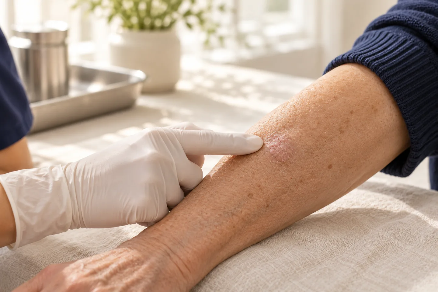
For people living in sunny climates such as Thailand—or expats and international patients who have accumulated substantial sun exposure over many years—persistent rough or changing lesions on the face, scalp, ears, forearms, or hands deserve proper assessment.
The key principle is simple: diagnosis comes before destruction. A lesion that looks like actinic keratosis is not always an actinic keratosis. If a spot is unusually thick, rapidly growing, painful, bleeding, ulcerated, or otherwise suspicious, a dermatologist may recommend a skin biopsy rather than simply freezing or treating it empirically.
What Is Actinic Keratosis?
Actinic keratosis is an area of abnormal keratinocyte growth in sun-damaged skin. It usually develops after years of cumulative exposure to ultraviolet radiation.
It is often described as a precancerous lesion because it lies on a biological spectrum between chronically sun-damaged skin and cutaneous squamous cell carcinoma.
Not every actinic keratosis will become skin cancer. Some remain stable and some may even regress. However, it is difficult to predict with certainty which individual lesion will eventually progress. Patients with actinic keratoses also tend to have significant cumulative sun damage and an increased overall risk of developing other skin cancers.
This is why AK should not simply be dismissed as a cosmetic sign of aging.
What Does Actinic Keratosis Look and Feel Like?
Actinic keratoses vary considerably in appearance.
A typical lesion may be:
- Rough or sandpaper-like
- Scaly or crusted
- Pink, red, skin-colored, yellowish, or brown
- Flat or slightly raised
- Easier to feel than to see
- Tender or sensitive when touched
- Persistent despite moisturizer
- Recurrent after the scale or crust is picked off
Some become considerably thicker and develop a hard keratin surface.
Common Locations
Actinic keratoses occur mainly on areas that have received substantial sun exposure, particularly:
- Forehead
- Cheeks
- Nose
- Temples
- Ears
- Bald or thinning scalp
- Lower lip
- Forearms
- Backs of the hands
A related condition occurring on the lip is called actinic cheilitis, most commonly affecting the lower lip.
What Causes Actinic Keratosis?
The main cause is cumulative ultraviolet radiation, particularly UV exposure from sunlight.
UV radiation damages DNA within skin cells. Over time, repeated damage can result in abnormal keratinocytes—the cells that make up most of the outer layer of the skin.
Risk therefore accumulates over many years rather than resulting from a single day in the sun.
Who Is at Higher Risk?
Risk is generally higher in people with:
- Fair skin
- A history of frequent sunburn
- Significant lifetime sun exposure
- Outdoor occupations or hobbies
- Older age
- Bald or thinning scalp
- Previous actinic keratoses
- Previous skin cancer
- A weakened immune system
- Organ transplantation or immunosuppressive treatment
Living for many years in tropical or subtropical environments can also contribute to cumulative UV exposure. Prolonged residence in tropical or subtropical climates is recognized as a relevant setting for AK development.
For patients living in Bangkok or elsewhere in Thailand, sun protection remains important even when most daily exposure occurs during ordinary activities rather than deliberate sunbathing.
Is Actinic Keratosis Skin Cancer?
Not exactly.
Actinic keratosis is generally classified as a precancerous lesion, rather than invasive skin cancer.
However, the distinction matters because AK and squamous cell carcinoma exist along a spectrum of abnormal keratinocyte growth.
Some actinic keratoses can progress to squamous cell carcinoma, a type of skin cancer that can invade deeper tissue and, in higher-risk cases, spread beyond the skin.
At the same time, it would be misleading to tell every patient that an individual AK will become cancer. Most individual lesions do not progress to invasive SCC, and predicting the behavior of one particular lesion is difficult.
The practical goal is therefore to identify suspicious lesions, treat appropriate AKs, monitor sun-damaged skin, and recognize skin cancer early.
Actinic Keratosis vs Squamous Cell Carcinoma
This distinction is one of the most important parts of evaluating a suspected AK.
An ordinary thin actinic keratosis may feel rough and remain relatively stable.
A lesion deserves greater concern when it becomes:
- Progressively thicker
- Firm or indurated
- Rapidly enlarging
- Persistently painful or tender
- Ulcerated
- Recurrently bleeding
- Non-healing
- Horn-like
- Significantly different from the patient’s other lesions
Tender, thickened, ulcerated, or enlarging AK-like lesions warrant particular attention because these features can raise concern for progression to SCC.
These changes do not automatically mean cancer, but they are reasons for dermatologic assessment rather than continued self-treatment.
Not Every Rough Spot Is Actinic Keratosis
Several skin conditions can resemble an actinic keratosis.
Possible alternatives include:
- Seborrheic keratosis
- Eczema
- Psoriasis
- Superficial fungal infection
- Viral wart
- Irritated benign growth
- Bowen disease or SCC in situ
- Invasive squamous cell carcinoma
- Superficial basal cell carcinoma
- Pigmented lesions that require assessment for melanoma
This is particularly important when a lesion is pigmented.
A brown or irregular facial lesion should not automatically be assumed to be a sun spot or actinic keratosis and treated destructively before the diagnosis is reasonably established.
How Does a Dermatologist Diagnose Actinic Keratosis?
Diagnosis usually begins with a careful skin examination.
A dermatologist will typically assess:
- Location
- Surface texture
- Thickness
- Scale or crust
- Color
- Borders
- Tenderness
- Duration
- Changes over time
- Previous treatment
- Degree of surrounding sun damage
Many typical actinic keratoses can be diagnosed clinically.
Dermoscopy
A dermatologist may also examine the lesion with dermoscopy, a magnified examination technique that reveals structures that are difficult to appreciate with the naked eye.
Dermoscopy can support the clinical diagnosis and help assess whether another diagnosis should be considered.
It does not eliminate the need for biopsy when a lesion remains suspicious.
When Is a Skin Biopsy Needed?
Not every actinic keratosis requires biopsy.
A biopsy may be appropriate when:
- The diagnosis is uncertain
- The lesion is unusually thick
- It is rapidly enlarging
- It repeatedly bleeds
- It is ulcerated
- It is persistently painful or tender
- There is concern for invasive SCC
- A treated lesion repeatedly returns
- The lesion fails to respond as expected
- The appearance is atypical
A biopsy may be required to exclude squamous cell carcinoma or when a presumed AK fails treatment.
What Happens During a Biopsy?
The area is usually numbed with local anesthetic. A small sample—or sometimes the entire lesion—is removed and sent for histopathological examination.
A pathologist examines the tissue under a microscope to determine what type of abnormal cells are present and whether the lesion is precancerous or cancerous.
This information can significantly change the treatment plan.
Treatment Options for Actinic Keratosis
There is no single treatment that is best for every actinic keratosis.
Treatment depends on:
- Number of lesions
- Thickness
- Location
- Size
- Whether lesions are isolated or widespread
- Previous treatment
- History of skin cancer
- Immune status
- Cosmetic considerations
- Ability to tolerate redness and downtime
Clinical guidelines strongly recommend UV protection, cryosurgery, topical 5-fluorouracil and topical imiquimod for AK, while photodynamic therapy and diclofenac receive conditional recommendations.
A useful way to understand treatment is to separate lesion-directed treatment from field treatment.
1. Cryotherapy with Liquid Nitrogen
Cryotherapy is one of the most commonly used treatments for individual actinic keratoses.
Liquid nitrogen is applied to the lesion to freeze and destroy abnormal cells.
It is particularly useful when there are only a few clearly defined lesions.
Advantages
- Quick procedure
- Usually performed during an outpatient visit
- No surgical incision
- Useful for isolated AKs
What Happens Afterwards?
The treated area may become:
- Red
- Swollen
- Tender
- Blistered
- Crusted
The crust then separates as the skin heals.
Limitations and Side Effects
Cryotherapy can occasionally cause:
- Prolonged redness
- Lighter skin color
- Darker pigmentation
- Scarring
- Incomplete clearance
- Recurrence
Longer or deeper freezing increases the possibility of pigment change or scarring.
For patients with Asian skin, post-treatment pigment changes can be particularly noticeable and should be discussed before treatment.
2. Topical 5-Fluorouracil (5-FU)
5-fluorouracil, commonly abbreviated as 5-FU, is a prescription medication applied to sun-damaged skin.
Unlike cryotherapy, which usually targets individual visible lesions, 5-FU can be used as field treatment.
This means the medication treats a broader area containing both visible AKs and abnormal sun-damaged cells that may not yet be obvious.
What Should Patients Expect?
Treatment intentionally produces inflammation.
During treatment the skin may become:
- Red
- Inflamed
- Crusted
- Sore
- Sensitive
The reaction can look dramatic, particularly when a larger facial area is treated.
This does not necessarily mean the medication is causing an allergy. An inflammatory response is an expected part of treatment.
However, the treatment schedule should be prescribed individually, and patients should know when the reaction is excessive enough to contact their dermatologist.
3. Imiquimod
Imiquimod is another prescription topical treatment that can be used for actinic keratoses.
Rather than directly freezing or removing the lesion, imiquimod modifies the local immune response so that abnormal cells are targeted.
It can be useful when treating an area containing multiple AKs.
Expected Reaction
Redness, inflammation, crusting, soreness and irritation can occur during treatment.
Treatment schedules vary according to formulation, location and clinical circumstances. Patients should therefore follow the prescribed regimen rather than copying a schedule found online.
4. Photodynamic Therapy
Photodynamic therapy (PDT) combines a photosensitizing medication with a specific light source.
The medication is applied to the affected skin and preferentially affects abnormal cells. Light activation then produces a reaction that damages those cells.
PDT can be useful for multiple actinic keratoses, particularly across a broader sun-damaged area.
Advantages
- Treats multiple lesions simultaneously
- Can address field damage
- May provide a useful cosmetic outcome in selected patients
Limitations
Treatment can cause:
- Pain or burning during illumination
- Redness
- Swelling
- Crusting
- Photosensitivity
- Temporary pigment changes
Availability and treatment protocols vary between medical facilities.
5. Curettage, Shave Removal or Electrosurgery
A thick or hyperkeratotic lesion may require physical removal rather than treatment with cream alone.
Options can include:
- Shave removal
- Curettage
- Electrosurgery
An important advantage is that removed tissue may be sent for histopathology when clinically appropriate.
This can be particularly useful when a thick lesion raises concern that it may represent something more advanced than a routine AK.
Curettage and electrocautery may be required for hypertrophic AKs or cutaneous horns.
6. Surgical Excision
Surgical excision is not required for most ordinary actinic keratoses.
It may become appropriate when:
- The diagnosis is uncertain
- Skin cancer is suspected
- Histopathology shows SCC
- The lesion has features requiring complete removal
The lesion is removed surgically and examined by a pathologist.
The major advantage is the ability to obtain tissue for diagnosis and assess the removed lesion. The trade-off is a permanent surgical scar.
Other Topical Treatments
Other prescription treatments are available in some countries, including diclofenac and tirbanibulin.
Diclofenac has a conditional recommendation in clinical guidelines, while treatment availability and approved indications can vary between countries.
Tirbanibulin is another short-course topical option used for AK of the face or scalp in some jurisdictions.
Patients in Thailand should discuss which medications are currently registered, available, and appropriate rather than assuming that every treatment described on an overseas website is locally available.
Individual Lesion Treatment vs Field Treatment
This distinction is particularly useful for patients with significant sun damage.
Lesion-Directed Treatment
This approach targets one or several visible lesions.
Examples include:
- Cryotherapy
- Curettage
- Shave removal
- Excision
It is often appropriate when there are only a few discrete AKs.
Field Treatment
Sometimes the visible AKs are only the most obvious signs of a much larger area of UV-damaged skin.
This is called field cancerization or field change.
The surrounding skin may contain abnormal keratinocytes even though individual lesions are not yet easily visible.
Field-directed treatments may include:
- 5-FU
- Imiquimod
- Photodynamic therapy
- Other appropriate topical treatments
The best approach may involve both methods—for example, treating a particularly thick lesion individually while addressing surrounding field damage separately.
Why You Should Not Simply Laser or Freeze Every Suspicious Spot
A destructive procedure removes or destroys tissue.
That can be useful when the diagnosis is sufficiently clear.
But if a suspicious lesion is destroyed before adequate assessment, there may be no tissue left for the pathologist to examine.
This matters because early SCC, SCC in situ, superficial basal cell carcinoma and other lesions can sometimes resemble benign or precancerous conditions.
For a lesion with concerning features, obtaining a diagnosis may be more important than immediately removing its visible surface.
This principle is especially important for recurrent, growing, bleeding, ulcerated, indurated or otherwise atypical lesions.
What Happens If a Biopsy Shows Squamous Cell Carcinoma?
At that point, the condition is no longer treated simply as an actinic keratosis.
Treatment depends on factors such as:
- Whether the SCC is in situ or invasive
- Tumor size
- Depth
- Location
- Histological features
- Immune status
- Whether it is a primary or recurrent tumor
Surgical excision is an important treatment for invasive cutaneous SCC. Selected higher-risk or anatomically sensitive tumors may require more specialized approaches such as Mohs micrographic surgery.
The appropriate treatment therefore depends on the pathology rather than simply on what the lesion looked like before biopsy.
Recovery After Actinic Keratosis Treatment
Recovery varies substantially according to the treatment.
Cryotherapy may produce a blister or crust followed by healing.
Field treatment with 5-FU or imiquimod can cause considerably more widespread redness, crusting and inflammation because a larger area is being treated.
Procedures such as curettage or excision create a wound that needs time to heal.
Your dermatologist should explain:
- How to clean the treated area
- Whether an ointment or dressing is required
- What degree of redness is expected
- Whether sun avoidance is necessary
- When makeup or skincare can be resumed
- When follow-up is needed
Can Actinic Keratosis Come Back?
Yes.
A successfully treated lesion may recur, and new AKs can develop elsewhere because treatment cannot erase the cumulative UV damage already present throughout the skin.
Patients who develop actinic keratoses therefore often benefit from continued surveillance rather than viewing treatment as a one-time event.
AKs can recur months or years after treatment, and people with AKs have an increased risk of additional keratinocyte cancers and other skin cancers.
Preventing New Actinic Keratoses
Sun protection is a core part of AK management and receives a strong recommendation in clinical guidelines.
Useful measures include:
- Broad-spectrum sunscreen
- Protective clothing
- Hats that adequately shade the face, scalp and ears
- Seeking shade when practical
- Avoiding deliberate tanning
- Reapplying sunscreen when appropriate
- Protecting commonly forgotten areas such as the ears, scalp, neck and backs of the hands
For patients living in Thailand, sun protection should be considered an everyday habit rather than something reserved for beach holidays.
Sun protection cannot reverse all previous UV damage, but it can reduce additional exposure.
When Should You See a Dermatologist?
Consider arranging a dermatology assessment if you notice a spot that:
- Persists for weeks or months
- Repeatedly becomes rough or crusted
- Grows progressively
- Becomes thicker
- Bleeds without clear injury
- Does not heal
- Develops an ulcer
- Becomes persistently painful or tender
- Changes noticeably in color or shape
- Returns after previous treatment
You should also consider periodic skin examination if you have extensive sun damage, numerous AKs, previous skin cancer, or significant immunosuppression.
What to Expect During an Actinic Keratosis Consultation in Bangkok
A dermatology consultation should focus first on establishing the diagnosis.
The dermatologist may ask about:
- How long the lesion has been present
- Whether it has changed
- Bleeding, pain or tenderness
- Previous treatments
- Previous skin cancers
- Sun exposure
- Medications
- Conditions affecting the immune system
The lesion and surrounding skin are then examined. Dermoscopy may be used when appropriate.
If the lesion appears typical, treatment options can be discussed according to its number, location and thickness.
If the diagnosis is uncertain or skin cancer is suspected, biopsy may be recommended before definitive treatment.
For patients with multiple sun-damaged areas, the consultation may also include discussion about broader skin surveillance rather than focusing only on the single spot that prompted the visit.
Frequently asked questions
Is actinic keratosis cancer?
Actinic keratosis is generally considered a precancerous lesion rather than invasive skin cancer. However, some AKs can progress to cutaneous squamous cell carcinoma.
Does every actinic keratosis need to be removed?
Treatment is commonly recommended, but management should be individualized. Factors include lesion appearance, symptoms, number of lesions, immune status, age, medical conditions and ability to tolerate treatment.
Can I tell an actinic keratosis from skin cancer by looking at it?
Not reliably in every case. Early SCC and other skin lesions can resemble AK. Dermatologic examination and sometimes biopsy are needed when the diagnosis is uncertain.
Should a thick actinic keratosis be biopsied?
A thick, tender, enlarging, ulcerated, bleeding or otherwise atypical lesion deserves careful evaluation. Biopsy may be needed to exclude SCC.
Is cryotherapy the best treatment for actinic keratosis?
Cryotherapy is very useful for isolated lesions, but it is not automatically the best option for every patient. Multiple AKs or widespread field damage may be better addressed with field treatment.
Is liquid nitrogen treatment painful?
Patients commonly experience brief stinging or burning during and shortly after freezing. A blister or crust can develop afterward.
Can actinic keratosis be treated with cream?
Yes. Prescription treatments include 5-FU and imiquimod, among others. The appropriate medication and regimen depend on the lesion and patient.
Why does my skin look worse during 5-FU treatment?
5-FU intentionally causes inflammation in treated sun-damaged skin. Redness and crusting can therefore be expected. Patients should still contact their dermatologist if the reaction is unusually severe or concerning.
Can actinic keratosis return after treatment?
Yes. Treated lesions can recur, and new AKs may develop in other sun-damaged areas.
Can actinic keratosis disappear by itself?
Some lesions can regress, but this cannot reliably predict whether another lesion will persist or progress. Suspicious or changing lesions should still be assessed.
Can actinic keratosis occur in Asian skin?
Yes. Although risk is higher in fairer skin types, AK can occur in people of any ethnicity, particularly with substantial cumulative UV exposure or immunosuppression.
Should I have a full skin examination if I have actinic keratosis?
It may be appropriate, particularly if you have multiple AKs, extensive sun damage, a previous skin cancer or other risk factors. AK indicates significant cumulative UV damage and is associated with increased skin-cancer risk.
Is actinic keratosis contagious?
No. Actinic keratosis is related primarily to UV-induced cellular damage and is not contagious.
Can I treat actinic keratosis myself?
A persistent suspicious lesion should ideally be diagnosed before treatment. Self-treating it as a wart, fungal infection or sun spot may delay the diagnosis of skin cancer.
What happens if my actinic keratosis keeps coming back after freezing?
Recurrent lesions should be reassessed. Depending on the appearance, a dermatologist may recommend another treatment or biopsy to confirm the diagnosis.
Related conditions
-
Full Body Skin Examination Full Body Skin Examination A full body skin examination is a head-to-toe assessment of the skin performed to look for suspicious moles, abnormal growths, precancerous changes and signs of skin cancer.

-
Suspicious Mole Assessment Suspicious Mole Assessment A suspicious mole assessment is a focused dermatology evaluation of a mole or pigmented skin lesion that is new, changing, unusual, bleeding, itching, or simply looks different from your other moles.
-
Dermoscopy Dermoscopy Dermoscopy is a non-invasive examination technique that allows a dermatologist to inspect skin lesions with greater detail than is possible with the naked eye alone.

-
Skin Biopsy (การวินิจฉัย / Diagnosis) Skin Biopsy (การวินิจฉัย / Diagnosis) A skin biopsy is a minor medical procedure in which a dermatologist removes a small sample of skin so it can be examined under a microscope. It is one of the most important diagnostic tools in dermatology because many skin conditions cannot be confirmed by appearance alone.