Basal Cell Carcinoma
Basal cell carcinoma (BCC) is a common type of skin cancer that usually grows slowly and is most often found on sun-exposed skin. Although BCC rarely spreads to distant parts of the body, it can continue growing into surrounding skin and deeper structures if left untreated. Treatment is therefore recommended after the diagnosis is confirmed.
For most patients, surgery is the main treatment for basal cell carcinoma. The best treatment, however, depends on the tumor’s location, size, histological subtype, whether it is a first or recurrent cancer, and individual patient factors. Standard surgical excision, Mohs micrographic surgery, topical medications, radiation therapy, curettage and other treatments each have specific roles.
For Thai patients, expatriates, tourists, and international patients seeking skin cancer treatment in Bangkok, the important first step is not simply choosing a procedure. It is confirming what type of skin cancer is present and whether it has features that make it low or high risk.
What Is Basal Cell Carcinoma?
Basal cell carcinoma is a malignant tumor arising from cells associated with the basal layer of the skin.
It belongs to the group commonly called non-melanoma skin cancers and behaves very differently from melanoma.
BCC usually grows locally rather than spreading through the body. Nevertheless, untreated tumors can gradually destroy surrounding tissue and may become considerably more difficult to treat when they are large, recurrent, deeply infiltrative, or located in anatomically important areas.
This is especially relevant for tumors on areas such as the:
- Nose
- Eyelids
- Ears
- Lips
- Forehead
- Scalp
- Cheeks
- Other parts of the face
In these locations, treatment must consider both complete cancer removal and preservation of healthy tissue and function.
What Causes Basal Cell Carcinoma?
Long-term exposure to ultraviolet radiation is an important risk factor for BCC.
Ultraviolet exposure accumulates throughout life. This means a person’s skin cancer risk reflects not only where they live now, but also previous sun exposure, outdoor activities, occupations, childhood exposure, and previous residence in high-ultraviolet environments.
Other factors that can increase risk include:
- Fair skin that burns easily
- Significant cumulative sun exposure
- Previous basal cell carcinoma
- Previous skin cancers
- Increasing age
- Previous radiation exposure
- Certain inherited conditions
- Reduced immune function
Living in a sunny climate such as Thailand makes appropriate sun protection particularly relevant, but BCC can occur in people of any nationality and should not be dismissed solely because someone has darker skin.
What Does Basal Cell Carcinoma Look Like?
BCC does not have one single appearance.
It may appear as:
- A pearly or shiny bump
- A pink or reddish growth
- A persistent red patch
- A slowly enlarging lesion
- A sore that repeatedly heals and returns
- A lesion that bleeds easily
- A crusted area that does not disappear
- A scar-like patch
- A pigmented brown, blue-gray, or dark lesion
Small visible blood vessels may sometimes be present over the surface.
Clinical literature notes that basal and squamous cell carcinomas can present as non-healing sores or raised, shiny, pearly, scar-like, red, scaly, bleeding, or crusted lesions.
Does BCC Always Look Like a Pearly Bump?
No.
This is a common misconception.
Superficial BCC may resemble eczema or dermatitis. Pigmented BCC can resemble a mole. Infiltrative or morpheaform forms may resemble scar tissue.
This is one reason persistent or unusual lesions should be examined rather than diagnosed from photographs alone.
Warning Signs That Should Be Checked by a Dermatologist
Consider dermatological assessment if you notice:
- A sore that does not heal
- A lesion that repeatedly bleeds or crusts
- A pink or pearly bump that slowly enlarges
- A persistent red patch that does not respond as expected to treatment
- A scar-like area without a history of injury
- A new or changing pigmented lesion
- A growth on the nose, eyelid, ear, lip, or other facial area
- A lesion that returns after previous removal
- A previously diagnosed skin cancer that appears to be recurring
Not every persistent lesion is cancer. Benign growths and inflammatory skin conditions can sometimes resemble BCC, which is why appropriate diagnosis matters before treatment.
How Is Basal Cell Carcinoma Diagnosed?
Diagnosis usually begins with a dermatological examination.
The dermatologist considers:
- Appearance
- Location
- Size
- Borders
- Duration
- Growth pattern
- Bleeding or ulceration
- Previous treatments
- Previous skin cancer history
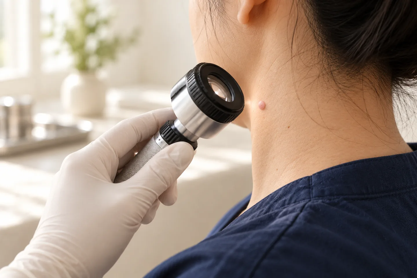
A dermatoscope may also be used to examine structures below the surface that are difficult to see with the naked eye.
Dermoscopic findings associated with BCC can include characteristic vascular and pigmented structures, but histopathology remains central to confirming the diagnosis and determining the histological subtype.
Is a Skin Biopsy Needed Before BCC Treatment?
A biopsy is commonly used to confirm suspected BCC and determine its histological subtype.
Possible techniques include:
- Shave biopsy
- Punch biopsy
- Incisional biopsy
- Excisional biopsy
Which technique is appropriate depends on the lesion.
After removal, the tissue is sent for histopathology, where a pathologist examines it under a microscope.
The pathology result can answer important questions such as:
- Is this actually basal cell carcinoma?
- What subtype is present?
- Are there aggressive growth characteristics?
- If the lesion was excised, are the margins clear?
Diagnosis and treatment should therefore be considered as part of the same pathway rather than treating a suspicious lesion without knowing what it is.
Why Does the Histological Subtype of BCC Matter?
Not all basal cell carcinomas behave identically.
Some tumors have relatively well-defined growth patterns, while others grow as small strands or nests extending beyond the visible edge.
Examples of patterns encountered in BCC include:
- Superficial
- Nodular
- Infiltrative
- Micronodular
- Morpheaform
Infiltrative, micronodular and morpheaform patterns are among features associated with greater risk of recurrence and can require a different surgical approach from a small, clearly defined superficial BCC.
This is why a treatment that is reasonable for one BCC may be inappropriate for another.
Low-Risk vs High-Risk Basal Cell Carcinoma
Treatment planning involves more than measuring the lesion.
Dermatologists consider a combination of factors.
Features That May Influence Risk Include
- Tumor location
- Tumor size
- Histological subtype
- Whether the borders are well defined
- Whether the tumor has previously been treated
- Whether it has recurred
- Depth or extent of invasion
- Patient immune status
- Other clinical and pathological features
Tumors on certain parts of the face require particular consideration because even relatively small cancers can involve functionally and cosmetically important structures.
Professional standards recommend risk stratification of BCC because treatment selection should reflect the clinical characteristics of the tumor.
Basal Cell Carcinoma Treatment Options
There is no single treatment that is best for every basal cell carcinoma.
The major options include:
- Standard surgical excision
- Mohs micrographic surgery
- Curettage and electrodesiccation in selected tumors
- Topical medication for selected superficial tumors
- Radiation therapy
- Photodynamic therapy in selected situations
- Cryosurgery in selected situations
- Systemic targeted therapy for certain advanced tumors
Surgery remains the cornerstone of BCC treatment, while nonsurgical approaches generally have more restricted indications.
Surgical Excision for Basal Cell Carcinoma
Standard surgical excision is one of the most commonly used treatments for BCC.
The procedure involves removing:
The visible cancer + an appropriate margin of apparently normal surrounding skin
The specimen is then sent to pathology.
The pathologist examines the tissue and assesses whether tumor is present at the edges of the specimen.
Advantages of Surgical Excision
Surgical excision provides several important benefits:
- Removes the tumor
- Provides tissue for histopathological examination
- Allows assessment of surgical margins
- Can be used for many different BCC subtypes
- Often provides definitive treatment in a single surgical episode
Surgery is particularly important for tumors where tissue confirmation and margin assessment are necessary.
Limitations of Surgical Excision
Possible disadvantages include:
- Scar formation
- Bleeding
- Infection
- Wound separation
- Need for sutures
- Need for a flap or skin graft for larger defects
- Possible need for further surgery if margins are involved
The cosmetic outcome depends on tumor size, location, wound-repair method and individual healing.
What Is a Surgical Margin?
A surgical margin is an area of clinically normal-looking skin removed around the visible tumor.
BCC cells may sometimes extend microscopically beyond what can be seen from the surface.
The purpose of the margin is therefore to remove microscopic extensions surrounding the visible cancer.
The appropriate margin is not identical for every tumor. It depends on the tumor’s risk characteristics.
What Does “Clear Margin” Mean?
After excision, the pathologist examines the tissue edges.
A report may describe margins as:
- Clear
- Negative
- Free of tumor
This generally means tumor cells were not identified at the examined specimen edge.
However, the significance of a margin result must be interpreted in the context of the tumor and surgical technique.
What Does “Positive Margin” or “Involved Margin” Mean?
An involved margin means tumor cells reach the edge of the tissue specimen.
This raises concern that microscopic BCC may remain in the skin.
Further treatment is often considered, which may include another excision or Mohs surgery depending on tumor characteristics and location. Inadequately excised high-risk tumors warrant particular attention.
What Is Mohs Surgery?
Mohs micrographic surgery is a specialized technique for removing skin cancer while examining the surgical margins during the procedure.
Instead of removing the tumor and waiting for routine permanent pathology, tissue is removed in stages and mapped.
The margins are examined microscopically. If residual tumor remains in a particular area, additional tissue can be removed specifically from that location.
The process continues until the examined margins are free of tumor.
When Is Mohs Surgery Considered for Basal Cell Carcinoma?
Mohs surgery can be particularly valuable when preserving healthy tissue is important or when the tumor has a higher risk of recurrence.
Situations in which it may be considered include selected:
- Facial BCCs
- Nasal tumors
- Eyelid tumors
- Ear tumors
- Lip-area tumors
- Recurrent BCCs
- Tumors with poorly defined borders
- Aggressive histological subtypes
- Tumors in locations where tissue preservation is important
The decision is based on the individual tumor rather than simply whether it is located on the face.
Mohs Surgery vs Standard Excision
Patients frequently ask which procedure is better.
The answer depends on the cancer.
| Standard Excision | Mohs Surgery |
|---|---|
| Tumor and planned surrounding margin removed | Tumor removed in mapped stages |
| Tissue usually assessed with routine pathology | Margins examined during the procedure |
| Appropriate for many BCCs | Particularly useful for selected high-risk tumors |
| Straightforward for many well-defined lesions | Maximizes margin assessment while conserving uninvolved tissue |
| Further surgery may be required if margins are involved | Additional involved areas can be removed during the same Mohs procedure |
Mohs surgery is not automatically necessary for every BCC.
A small, low-risk BCC on the trunk may be managed very differently from an infiltrative recurrent tumor on the nose.
Is Mohs Surgery Better for Facial BCC?
For appropriately selected facial or high-risk tumors, Mohs surgery has important advantages because it combines detailed margin assessment with conservation of surrounding healthy tissue.
This can be particularly valuable around structures such as:
- Nose
- Eyelids
- Ears
- Lips
However, the treatment decision should still consider tumor subtype, size, exact location, recurrence status and individual patient factors.
What Happens After the Cancer Is Removed?
Removing the cancer leaves a surgical defect.
Depending on its size and location, the wound may be managed by:
- Direct closure with stitches
- Allowing the wound to heal naturally
- Local skin flap
- Skin graft
- Other reconstructive techniques
A larger wound does not necessarily mean that something went wrong. Skin cancer can extend beyond the part visible on the surface, and adequate removal sometimes requires a larger defect than patients initially expect.
Curettage and Electrodesiccation
This technique involves scraping the tumor tissue with a curette and treating the base using electrical energy.
It may be appropriate for selected low-risk BCCs, particularly certain lesions on the trunk or extremities.
Advantages
Relatively quick
Does not require a traditional elliptical excision
Useful in carefully selected low-risk lesions
Limitations
The surgeon cannot assess the entire microscopic margin in the same way as with margin-controlled surgery.
It is therefore not interchangeable with Mohs surgery or standard excision for all tumors.
Can Basal Cell Carcinoma Be Treated With Cream?
Selected superficial, low-risk basal cell carcinomas may sometimes be treated with topical medication.
Options can include:
- Imiquimod
- Topical fluorouracil
These treatments are not appropriate substitutes for surgery in every BCC.
Expert medical consensus notes that nonsurgical treatments generally have lower cure rates than surgical approaches and should primarily be considered for low-risk BCC or when surgery is contraindicated.
Advantages of Topical Treatment
In appropriate cases:
- No surgical incision
- Can treat a superficial area
- May be considered when surgery is unsuitable
Limitations
Potential disadvantages include:
- Treatment takes time
- Significant inflammation may occur
- Redness, crusting, erosion and discomfort can develop
- No conventional excision specimen is available afterward to confirm complete margin clearance
- Deeper tumor components may not be adequately treated
- Not appropriate for many higher-risk BCCs
The diagnosis and subtype should therefore be established appropriately before selecting topical treatment.
Can Imiquimod Treat Basal Cell Carcinoma?
Imiquimod can be used for selected superficial BCCs.
It works by stimulating a local immune response.
During treatment, patients may develop:
- Redness
- Inflammation
- Crusting
- Erosion
- Tenderness
These reactions can be expected as part of treatment, but severe reactions should be reviewed by the treating doctor.
Imiquimod should not be viewed as a universal alternative to surgery.
Can Fluorouracil Cream Treat Basal Cell Carcinoma?
Topical fluorouracil can be considered for selected superficial BCCs.
Its effect is mainly superficial, which is an important limitation. Deeper tumor components may persist despite apparent improvement at the surface.
This is why confirming the type and depth characteristics of the tumor before choosing topical treatment is important.
Can Basal Cell Carcinoma Be Frozen?
Cryosurgery destroys tissue by freezing it.
It can be used for selected BCCs, but it is not the preferred treatment for every tumor.
Important limitations include reduced ability to assess microscopic margins and difficulty knowing immediately whether every cancer cell has been destroyed.
It is therefore generally reserved for carefully selected cases rather than being used simply because freezing is convenient.
Can Laser Remove Basal Cell Carcinoma?
Laser treatment is not the standard first choice for most BCCs.
Professional guidelines list laser surgery among possible approaches but note that it is rarely used for basal cell carcinoma.
This distinction matters because patients sometimes assume that a visible skin cancer can simply be “lasered off.
”
When malignancy is suspected, destroying a lesion without appropriate diagnostic tissue or margin assessment may remove the visible surface while leaving uncertainty about deeper tumor.
Photodynamic Therapy for Basal Cell Carcinoma
Photodynamic therapy combines a photosensitizing medication with a specific light source.
It can be considered for selected superficial tumors.
Potential advantages include avoiding conventional surgery in certain situations.
Limitations include:
- Not appropriate for every BCC subtype
- Treatment-site inflammation
- Pain during or after treatment
- Photosensitivity precautions depending on the treatment
- Recurrence risk that must be considered when comparing it with surgical treatment
Treatment selection should be based on tumor risk rather than cosmetic preference alone.
Radiation Therapy for Basal Cell Carcinoma
Radiation therapy uses ionizing radiation to destroy cancer cells.
It can be considered when surgery is unsuitable, would be particularly difficult, or in selected recurrent or anatomically challenging tumors.
Current clinical guidelines note that radiation can be useful when surgery would require difficult or extensive reconstruction, including selected tumors around areas such as the nose or ears.
Advantages
Avoids surgical removal in selected patients
Can treat tumors in anatomically difficult locations
Useful when surgery is medically unsuitable
Limitations
Usually requires multiple treatment visits
Skin reactions can occur
Long-term pigment and vascular changes may occur
It does not provide a surgical specimen demonstrating complete removal
It is unsuitable in certain medical or genetic conditions
Radiation therapy is therefore an important option, but not automatically preferable to surgery.
What About Advanced Basal Cell Carcinoma?
Most BCCs are treated locally.
A small proportion become locally advanced, meaning the cancer has grown extensively and cannot reasonably be managed with standard local treatment.
Metastatic BCC is rare but can occur.
For advanced disease that cannot be managed with local treatment, systemic therapy may be considered.
Options can include drugs targeting the Hedgehog signaling pathway, such as:
- Vismodegib
- Sonidegib
Advanced cases require specialist multidisciplinary evaluation rather than routine treatment of a small skin lesion.
Which BCC Treatment Is Best?
There is no universally “best” procedure.
The appropriate treatment depends on questions such as:
Where is the tumor?
A lesion on the nose may require a different strategy from one on the back.
How large is it?
Larger tumors can require wider removal and more complicated reconstruction.
What subtype is it?
Superficial BCC behaves differently from infiltrative or morpheaform BCC.
Is it a first tumor or a recurrence?
Recurrent cancers often require more careful margin control.
Are the borders clearly visible?
Poorly defined tumors may extend beyond what is apparent clinically.
What does the pathology show?
Histology can significantly alter the treatment plan.
What is the patient’s overall health?
Age alone does not determine treatment. Medical conditions, medications, wound healing, treatment goals and ability to undergo procedures all matter.
Why Simply Removing the Visible Part May Not Be Enough
A skin cancer can extend microscopically beyond its visible border.
This is especially relevant for infiltrative growth patterns.
The visible lesion is therefore not necessarily the complete tumor.
Effective treatment aims to address both:
Visible cancer + possible microscopic extension
This is one reason margin assessment is important in surgical management.
Can Basal Cell Carcinoma Come Back After Treatment?
Yes.
No treatment can guarantee that BCC will never recur.
Risk of recurrence depends on factors including:
- Tumor subtype
- Location
- Size
- Previous treatment
- Margin status
- Treatment method
- Whether the tumor was recurrent before treatment
Recurrent BCC is associated particularly with previous incomplete excision, aggressive histological patterns, and head-and-neck location.
What Does Recurrent BCC Look Like?
Recurrence may appear as:
- A new bump within or next to a scar
- Persistent crusting
- Recurrent bleeding
- A non-healing area
- Progressive thickening
- A new ulcer
- A scar that begins changing
Changes within a previous skin-cancer treatment site should be assessed rather than assumed to be normal scar tissue.
If I Have Had One BCC, Can I Get Another?
Yes.
Having previously developed BCC indicates that your skin has already demonstrated susceptibility to skin cancer.
Current clinical guidelines recommend ongoing skin checks after a BCC diagnosis to look for recurrence and new primary skin cancers.
Follow-up should therefore consider both:
the original treatment site and the rest of the skin.
Follow-Up After Basal Cell Carcinoma Treatment
Follow-up may include:
- Examination of the surgical scar
- Checking for recurrence
- Examination for new suspicious lesions
- Review of sun-protection practices
- Advice about self-examination
- Full-body skin examination when appropriate
The exact follow-up schedule should be individualized according to the patient’s history and tumor risk.
Recovery After BCC Surgery
Recovery depends substantially on:
- Tumor size
- Location
- Type of surgery
- Wound closure
- Whether a flap or graft is required
- Individual healing characteristics
- Common temporary effects can include:
- Tenderness
- Swelling
- Bruising
- Mild bleeding
- Tightness around stitches
Every surgical procedure also creates some degree of scar.
What Will the Scar Look Like?
Scar appearance varies.
Important factors include:
- Location
- Size and depth of the surgical defect
- Direction of wound closure
- Skin tension
- Reconstruction technique
- Individual tendency to scar
- Infection or wound complications
Scars generally change over time and may become less noticeable as they mature.
For facial skin cancer, treatment planning often involves balancing two priorities:
complete tumor removal and preservation of normal tissue.
Cancer clearance should not be compromised simply to make the initial wound smaller.
Possible Complications of Skin Cancer Surgery
Potential complications include:
- Bleeding
- Infection
- Bruising
- Swelling
- Wound separation
- Delayed healing
- Scar formation
- Pigment change
- Numbness around the scar
- Need for additional surgery
- Recurrence
The exact risk depends on the procedure and tumor location.
How Should I Care for the Wound After Surgery?
Follow the wound-care instructions given by the treating clinician.
Depending on the procedure, this may involve:
- Keeping the wound clean
- Applying the recommended dressing
- Avoiding unnecessary friction or trauma
- Limiting activities that pull on the wound
- Returning for wound review
- Returning for suture removal when required
- Seek medical review if there is:
- Persistent significant bleeding
- Increasing pain
- Increasing redness or swelling
- Pus
- Fever with worsening wound symptoms
- Wound separation
Can I Fly After Basal Cell Carcinoma Surgery?
Many patients receiving treatment in Bangkok are tourists or international patients.
Whether flying is appropriate depends less on the diagnosis itself and more on:
- Size and location of surgery
- Type of reconstruction
- Risk of bleeding
- Need for dressing changes
- Timing of suture removal
- Need to receive the pathology result
- Possibility of further surgery
International patients should discuss travel plans before surgery so that pathology review and postoperative care can be coordinated.
Can I Have BCC Removed in Bangkok and Follow Up in My Home Country?
Potentially, yes, provided continuity of care is planned carefully.
Before leaving Thailand, patients should ideally know how they will obtain:
- Pathology report
- Surgical report when applicable
- Margin status
- Wound-care instructions
- Suture-removal instructions
- Recommendations for additional treatment
- Follow-up plan
If further treatment is required, having complete medical documentation makes transfer of care much easier.
Should a Tourist Wait Until Returning Home for BCC Treatment?
There is no single answer.
Factors to consider include:
- Whether the diagnosis is already confirmed
- Tumor location
- Tumor size
- Growth rate
- Histological subtype
- Whether the lesion is recurrent
- How long the patient will remain in Thailand
- Whether adequate postoperative follow-up can be arranged
A small BCC is usually not the same type of immediate emergency as a rapidly progressing systemic illness, but unnecessary delay can allow continued local growth.
A dermatologist can help determine how urgently treatment should be arranged.
Is Basal Cell Carcinoma Dangerous?
BCC usually behaves differently from melanoma.
It rarely spreads to distant organs, but “rarely spreads” should not be confused with “harmless.
”
Untreated BCC can progressively invade:
- Skin
- Subcutaneous tissue
- Cartilage
- Other nearby structures
Large, neglected or recurrent tumors can become substantially more difficult to manage.
Does BCC Need Treatment If It Does Not Hurt?
Yes, absence of pain does not mean the tumor is harmless.
Many BCCs are painless.
Treatment decisions are based on the diagnosis and tumor characteristics, not whether the lesion hurts.
Can Basal Cell Carcinoma Disappear by Itself?
A suspected or biopsy-confirmed BCC should not be assumed to resolve permanently without treatment.
A lesion may temporarily appear smaller, less inflamed, or crust over and heal superficially while tumor remains.
A biopsy can also remove a substantial visible portion of a small lesion, but this does not automatically prove that no microscopic cancer remains.
What If the BCC Seems Gone After Biopsy?
This is a particularly important question.
After biopsy, the visible lesion may appear to disappear completely.
However, microscopic tumor may still remain beneath or around the biopsy site.
Treatment decisions should therefore be based on:
- Pathology diagnosis
- Type of biopsy
- Margin information when available
- Original tumor characteristics
- Clinical examination
Appearance alone after biopsy is not sufficient to establish complete tumor clearance.
Can BCC Be Mistaken for a Benign Skin Lesion?
Yes.
BCC can resemble several benign conditions, including:
- Seborrheic keratosis
- Benign mole
- Scar
- Eczema
- Inflamed follicle
- Other non-cancerous growths
Conversely, a lesion that looks alarming may turn out to be benign.
This is why suspicious lesions should be diagnosed appropriately rather than automatically treated as cancer—or automatically dismissed as harmless.
Basal Cell Carcinoma vs Melanoma
These are different skin cancers.
Basal Cell Carcinoma
Usually:
- Grows locally
- Often progresses slowly
- Rarely metastasizes
- Commonly occurs on sun-exposed skin
Melanoma
Melanoma arises from melanocytes, the cells responsible for pigment.
It has a greater potential to invade and spread to other parts of the body and requires a different diagnostic, staging and treatment pathway.
A pigmented lesion should therefore not automatically be assumed to be BCC.
Basal Cell Carcinoma vs Squamous Cell Carcinoma
BCC and squamous cell carcinoma are both keratinocyte skin cancers, but they behave differently.
Squamous cell carcinoma has a greater potential for metastatic spread than typical BCC and has its own risk-assessment and treatment considerations.
Histopathology distinguishes the two and provides information needed for appropriate management.
Can You Prevent Another Basal Cell Carcinoma?
Not every BCC can be prevented, but reducing ultraviolet exposure is an important part of prevention.
Practical measures include:
- Seeking shade when ultraviolet exposure is intense
- Wearing protective clothing
- Wearing a hat when appropriate
- Using broad-spectrum sunscreen
- Reapplying sunscreen when necessary
- Avoiding intentional tanning
- Checking your own skin for new or changing lesions
- Attending follow-up examinations when recommended
For people living in Thailand, sun protection is relevant throughout the year rather than only during holidays.
What to Expect During a BCC Consultation in Bangkok
A dermatology consultation for suspected or confirmed basal cell carcinoma may involve:
Review of the Lesion
The doctor assesses:
- Location
- Size
- Borders
- Appearance
- Ulceration
- Previous treatment
Medical History
Important information may include:
- Previous skin cancers
- Previous pathology reports
- Previous surgery
- Immune-suppressing conditions or medications
- Blood-thinning medications
- Relevant medical conditions
Dermoscopic Examination
A dermatoscope may be used when appropriate.
Biopsy or Pathology Review
If the diagnosis has not been established, biopsy may be recommended.
If the patient already has a pathology result, this should be reviewed before treatment planning.
Treatment Discussion
The doctor can then discuss whether the lesion is more appropriately managed with:
- Standard excision
- Mohs surgery
- Another selected treatment
- Referral for more specialized treatment
For international patients, travel timing and continuity of postoperative care should also form part of the discussion.
What Should International Patients Bring to a Skin Cancer Consultation?
If you have already been evaluated elsewhere, bring whatever is available:
- Pathology report
- Previous biopsy report
- Operative report
- Clinical photographs
- Dermoscopic photographs
- Medication list
- Previous skin cancer history
For complex or uncertain pathology, the original pathology slides or tissue blocks may sometimes be relevant for specialist review.
Questions to Ask Before Basal Cell Carcinoma Treatment
Useful questions include:
Has the diagnosis been confirmed by pathology?
What histological subtype do I have?
Is this considered a lower-risk or higher-risk BCC?
Why is this particular treatment recommended?
Is standard excision appropriate?
Should Mohs surgery be considered?
Will the removed tissue be examined pathologically?
How will the margins be assessed?
What happens if the margins are involved?
How will the wound be repaired?
What scar should I expect?
When will I receive the final pathology result?
Will I need further treatment?
When should I have another skin examination?
Can follow-up be continued in another country?
Frequently asked questions
What is the best treatment for basal cell carcinoma?
There is no single best treatment for every BCC. Surgery is the mainstay of treatment, but the appropriate method depends on tumor location, size, subtype, recurrence status and patient factors.
Does basal cell carcinoma need to be removed?
A confirmed BCC generally requires treatment because it can continue to grow and invade surrounding tissue.
Is basal cell carcinoma life-threatening?
Most BCCs do not spread to distant organs, but neglected or advanced tumors can cause substantial local destruction. Metastatic disease is rare.
Can BCC spread to other organs?
It can, but metastatic BCC is rare.
Can basal cell carcinoma grow into bone?
Advanced, neglected tumors can invade deeply into surrounding structures.
Does BCC hurt?
It may be painless. Pain is not required for the diagnosis.
Does BCC bleed?
It can. Recurrent bleeding or crusting is one possible presentation.
Does BCC heal and come back?
Some lesions can crust, appear to heal, and then break down or bleed again.
Do I need a biopsy before treatment?
A biopsy is commonly used to confirm the diagnosis and identify the histological subtype before definitive treatment.
Can BCC be diagnosed with dermoscopy?
Dermoscopy can provide valuable diagnostic clues, but histopathology is generally used to confirm the diagnosis and subtype.
What is the difference between biopsy and excision?
A biopsy obtains tissue for diagnosis. An excision removes the tumor with a surrounding margin and can serve as definitive treatment in appropriate cases.
What is Mohs surgery?
Mohs surgery removes skin cancer in mapped stages while the margins are examined microscopically during the procedure.
Does every facial BCC need Mohs surgery?
No. Treatment depends on exact location, tumor size, subtype, border definition, recurrence status and other risk factors.
When should Mohs surgery be considered?
It is particularly useful for selected high-risk tumors, recurrent BCCs and cancers in areas where detailed margin assessment and tissue preservation are important.
Is standard excision effective for BCC?
Standard surgical excision is an established treatment for many basal cell carcinomas and remains one of the main treatment approaches.
What happens to BCC after it is removed?
The specimen can be sent to pathology to confirm the diagnosis and assess relevant histological features and margins.
What does a positive margin mean?
It means tumor reaches an examined edge of the specimen, raising concern that residual cancer may remain.
What happens if the margins are positive?
Further treatment is often considered, such as re-excision or Mohs surgery depending on the tumor.
Can BCC be treated with cream?
Selected superficial, low-risk BCCs can sometimes be treated with topical medication.
Can imiquimod treat BCC?
Yes, in appropriately selected superficial BCCs, but it is not appropriate for every tumor.
Can fluorouracil treat BCC?
Topical fluorouracil can be used for selected superficial BCCs, but its superficial effect limits its usefulness for deeper disease.
Is cream treatment as effective as surgery?
Nonsurgical treatments generally have lower cure rates than surgical treatment and should be reserved for appropriate clinical situations.
Can BCC be frozen?
Cryosurgery can be used for selected lesions but is not appropriate for all BCCs.
Can BCC be removed with laser?
Laser surgery is rarely used as a treatment for BCC and is not the standard first-line approach for most tumors.
Can radiation treat BCC?
Yes. Radiation therapy can be useful when surgery is unsuitable or in selected anatomically difficult or recurrent cases.
Does BCC come back after surgery?
It can. Recurrence risk depends on tumor and treatment characteristics.
Can BCC recur after clear margins?
Recurrence is still possible, although margin status is an important factor in assessing treatment completeness and future risk.
Can I develop another BCC somewhere else?
Yes. Patients who have developed BCC have an increased risk of developing additional skin cancers and should continue skin surveillance.
How often should I have my skin checked after BCC?
Follow-up should be individualized. Clinical guidelines recommend at least annual assessment after BCC for recurrence or new primary skin cancers.
Does BCC surgery leave a scar?
Yes. Any surgery that cuts through the skin produces a scar, although its final appearance varies.
How long does BCC surgery take to heal?
Healing time depends on the tumor location, wound size and reconstruction method, so there is no single recovery period appropriate for every patient.
Can I exercise after BCC surgery?
Activity restrictions depend on the wound location and closure. Exercise that stretches or puts pressure on a fresh surgical wound may need to be temporarily limited.
Can I shower after BCC surgery?
Follow the specific wound-care instructions provided after the procedure because recommendations vary with the dressing and closure technique.
Can I fly after BCC surgery?
Often possible, but timing should consider wound care, bleeding risk, suture removal, pathology results and possible need for further treatment.
Can I have BCC treatment while visiting Bangkok?
Treatment during travel may be possible, but adequate time should be allowed for diagnosis, pathology, wound care and follow-up.
What if my BCC disappears after biopsy?
Residual microscopic tumor may remain even when nothing obvious is visible. Treatment decisions should be based on the pathology and clinical situation rather than appearance alone.
Can I wait to treat BCC?
The urgency varies by tumor, but confirmed BCC should not simply be ignored because continued growth can make treatment more difficult.
Is BCC the same as melanoma?
No. They arise from different cells and have very different biological behavior and treatment pathways.
Is BCC the same as squamous cell carcinoma?
No. Both are skin cancers, but their biological behavior and risk assessment differ.
Should I have a full-body skin examination after BCC?
Because a person with BCC is at risk of developing additional skin cancers, examination of the rest of the skin and ongoing surveillance can be appropriate.
What should I bring to a BCC consultation in Bangkok?
Bring previous pathology reports, biopsy or surgery reports, photographs, medication information and previous skin-cancer records when available.
Can pathology from another country be used in Thailand?
Previous pathology reports are useful. In selected cases, review of the original slides or tissue may also be considered before major treatment decisions.
Related conditions
-
Full Body Skin Examination Full Body Skin Examination A full body skin examination is a head-to-toe assessment of the skin performed to look for suspicious moles, abnormal growths, precancerous changes and signs of skin cancer.

-
Suspicious Mole Assessment Suspicious Mole Assessment A suspicious mole assessment is a focused dermatology evaluation of a mole or pigmented skin lesion that is new, changing, unusual, bleeding, itching, or simply looks different from your other moles.
-
Dermoscopy Dermoscopy Dermoscopy is a non-invasive examination technique that allows a dermatologist to inspect skin lesions with greater detail than is possible with the naked eye alone.

-
Skin Biopsy (การวินิจฉัย / Diagnosis) Skin Biopsy (การวินิจฉัย / Diagnosis) A skin biopsy is a minor medical procedure in which a dermatologist removes a small sample of skin so it can be examined under a microscope. It is one of the most important diagnostic tools in dermatology because many skin conditions cannot be confirmed by appearance alone.