






























Suspicious Mole Assessment
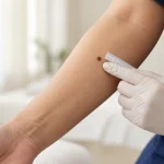
A suspicious mole assessment is a focused dermatology evaluation of a mole or pigmented skin lesion that is new, changing, unusual, bleeding, itching, or simply looks different from your other moles.

Most moles are benign. However, melanoma and some other skin cancers can initially resemble an ordinary mole. Important warning signs include a new lesion, a mole that changes in size, shape or color, or a spot that looks noticeably different from the rest of your moles.
During an assessment, a dermatologist usually examines the lesion clinically and may use dermoscopy, a magnified examination that reveals structures not easily visible to the naked eye. Depending on the findings, the mole may be considered benign, photographed and monitored, or biopsied for microscopic examination.
For Thai patients, expatriates, tourists and international patients in Bangkok, the most important message is simple: a suspicious mole does not automatically mean skin cancer, but a changing or unusual mole should not be ignored.
What Is a Suspicious Mole?
There is no single appearance that makes a mole suspicious.
A mole becomes more concerning when its appearance, behavior or history differs from what would normally be expected.
Features that may justify dermatological assessment include:
- A new mole or pigmented spot
- A mole that is changing
- Increasing asymmetry
- An irregular or poorly defined border
- Several colors within one lesion
- Rapid growth
- A mole that looks different from your other moles
- Recurrent bleeding
- Persistent crusting
- A new lump within a mole
- Persistent itching or tenderness
- A lesion that repeatedly breaks down
- New unexplained pigmentation beneath a nail
- A changing pigmented lesion on the palm or sole
Professional clinical guidance recommends medical evaluation of new, changing, itching or bleeding spots, while emphasizing new lesions, change and the “ugly duckling” sign as important melanoma warning features.
Does a Suspicious Mole Mean Melanoma?
No.
Many lesions that worry patients turn out to be benign.
A suspicious-looking spot may ultimately be:
- A normal mole
- An atypical mole
- Seborrheic keratosis
- Solar lentigo
- Angioma
- Irritated benign growth
- Pigmented basal cell carcinoma
- Another benign or inflammatory skin lesion
Conversely, some melanomas can initially look relatively ordinary.
This is why diagnosis should not rely only on whether a lesion “looks scary.
”
A dermatologist considers the mole’s appearance, history of change, the patient’s other moles, risk factors and, when appropriate, dermoscopic features.
What Does a Normal Mole Usually Look Like?
Typical benign moles are often:
- Symmetrical
- Round or oval
- Uniform in color
- Clearly bordered
- Stable over time
They may be flat or slightly raised.
However, benign moles do not all look identical. A person may naturally have moles of different sizes, shades and shapes.
The important question is often not simply:
“Does this mole look unusual?”
but also:
“Is this mole unusual for this particular person?”
Most ordinary moles remain relatively stable, although gradual changes can occur over many years.
What Does Melanoma Look Like?
Melanoma can appear in many different ways.
It may present as:
- A new dark spot
- A changing existing mole
- A multicolored lesion
- An irregularly shaped mole
- A rapidly enlarging lesion
- A black, brown, red or pink lesion
- A lesion with little or no dark pigment
- A dark streak beneath a nail
- A changing lesion on the sole
- A firm growing nodule
Some melanomas are relatively symmetrical. Some are smaller than 6 millimeters. Some are pink or red rather than brown or black.
Therefore, no checklist can identify every melanoma.
The ABCDE Warning Signs of Melanoma
The ABCDE method is a practical way to identify some suspicious pigmented lesions.
A — Asymmetry
One half of the mole does not resemble the other half.
B — Border
The border may be:
- Irregular
- Scalloped
- Notched
- Blurred
- Poorly defined
C — Color
Instead of one consistent color, a suspicious lesion may contain several shades, including:
- Tan
- Brown
- Black
- Red
- Pink
- White
- Blue
D — Diameter
Melanomas are often larger than 6 millimeters when diagnosed, but importantly, melanoma can be smaller than 6 millimeters. Size alone should never be used to rule out cancer.
E — Evolving
Change is one of the most important warning signs.
A lesion may change in:
- Size
- Shape
- Color
- Elevation
- Surface
- Symptoms
Clinical guidelines emphasize the ABCDE method while also noting that melanoma can be smaller than the traditional 6-millimeter threshold.
What Is the Ugly Duckling Sign?
The ugly duckling sign describes a mole that looks noticeably different from the person’s other moles.
For example, if nearly all of someone’s moles are:
- Small
- Light brown
- Round
- Uniform
but one mole is:
- Very dark
- Irregular
- Larger
- Multicolored
that lesion may deserve closer evaluation.
This concept is particularly useful because every person’s normal mole pattern is different.
A lesion does not necessarily need to be dramatic to be concerning. Sometimes being different from the rest is enough to justify examination.
Is a Changing Mole Always Cancer?
No.
Benign moles can change for several reasons.
Changes may occur with:
- Age
- Hormonal changes
- Pregnancy
- Irritation
- Repeated friction
- Trauma
- Inflammation
However, because evolution is also an important warning sign for melanoma, a meaningful unexplained change should be assessed rather than assumed to be harmless.
Is a New Mole in an Adult Suspicious?
Not every new adult mole is melanoma.
However, new pigmented lesions appearing later in adulthood deserve greater attention than longstanding stable moles, particularly when the new lesion looks different from existing moles or continues to change.
Clinical literature specifically lists the development of a new mole later in adult life among situations in which expert evaluation may be appropriate.
Does a Bleeding Mole Mean Cancer?
Not necessarily.
A raised benign mole can bleed if it is:
- Scratched
- Shaved
- Rubbed by clothing
- Caught on jewelry
However, spontaneous bleeding or repeated bleeding without an obvious injury should be assessed.
The same applies to a lesion that repeatedly crusts, breaks down or appears to heal and then returns.
Bleeding and surface changes are recognized warning features that can justify dermatological examination.
Is an Itchy Mole Dangerous?
Itching alone does not mean melanoma.
Common benign causes include:
- Dry skin
- Eczema
- Irritation
- Friction
- Insect bites
- Scratching
However, persistent itching in a mole that is also changing, bleeding, growing or becoming irregular warrants assessment.
Is a Painful Mole More Likely to Be Cancer?
Not necessarily.
Most melanomas are not painful.
Pain can occur for many benign reasons, particularly after irritation or trauma.
More importantly, lack of pain does not make a lesion safe.
A suspicious mole should be assessed according to its appearance and evolution rather than whether it hurts.
What About a Mole That Suddenly Becomes Raised?
A mole becoming more elevated can occur for benign reasons, but a rapidly developing bump or nodule deserves attention.
A particularly important limitation of the ABCDE rule is that nodular melanoma can sometimes be relatively symmetrical and uniformly colored, making it less obvious than the classic irregular melanoma.
A newly developing or rapidly growing firm nodule should therefore be examined even if it does not satisfy every ABCDE criterion.
What Is Amelanotic Melanoma?
Most people associate melanoma with a dark brown or black mole.
However, some melanomas contain very little visible pigment.
These are sometimes called amelanotic melanomas and may appear:
- Pink
- Red
- Flesh-colored
- Lightly pigmented
Because they do not resemble the classic dark melanoma, they can be more difficult to recognize.
Dermatological literature specifically notes that amelanotic melanoma may appear pink or red without the expected pigmentation.
Can Melanoma Occur on the Palm or Sole?
Yes.
Melanoma can develop on:
- Palms
- Soles
- Fingers
- Toes
- Beneath nails
These locations are particularly important in Asian and darker skin types because melanoma can occur in acral areas even when there has not been extensive sun exposure.
A changing dark spot on the sole should therefore not automatically be assumed to be a bruise, callus or pigmentation.
What About a Dark Line Under a Nail?
Many dark nail streaks are benign.
Possible causes include:
- Normal pigmentation
- Trauma
- Bleeding beneath the nail
- Benign melanocytic pigmentation
- Medication-related pigmentation
- Fungal or inflammatory nail disease
However, a new or changing pigmented band involving one nail may occasionally represent melanoma of the nail unit.
Features that deserve assessment include:
- A band becoming wider
- Increasingly irregular coloration
- New pigment in a single nail
- Pigment spreading onto surrounding skin
- Associated nail destruction
Clinical guidance notes that melanoma in harder-to-see areas may appear as a dark line or streak beneath a fingernail or toenail.
How Does a Dermatologist Assess a Suspicious Mole?
A high-quality mole assessment involves more than looking at the lesion for a few seconds.
The dermatologist usually considers three main elements:
- History
- Clinical examination
- Dermoscopic examination when appropriate
The decision about whether to observe, monitor or biopsy is based on the overall picture.
Medical History During a Suspicious Mole Assessment
The dermatologist may ask:
- When did you first notice the mole?
- Was it present during childhood?
- Has it recently changed?
- Has it grown?
- Has its color changed?
- Has it bled?
- Does it itch or hurt?
- Has it been repeatedly injured?
- Have you had melanoma before?
- Have you had basal cell or squamous cell carcinoma?
- Have you previously had atypical moles removed?
- Is there melanoma in your family?
- Have you experienced significant sun exposure?
- Have you had severe sunburns?
- Are you immunosuppressed?
A reliable history of change can be particularly important because a dermatologist sees the lesion only at one moment in time.
Clinical Examination
The dermatologist examines features such as:
- Size
- Symmetry
- Shape
- Border
- Color
- Surface
- Elevation
- Ulceration
- Crusting
- Bleeding
- Surrounding skin
The lesion may also be compared with other moles.
For some patients, examining the rest of the skin can reveal whether the suspicious mole truly differs from the person’s usual pattern.
What Is Dermoscopy?
Dermoscopy is a non-invasive method of examining skin lesions using a handheld magnifying device with specialized illumination.
It allows the dermatologist to see colors and structures beneath the outer skin surface that may not be visible to the naked eye.
Dermoscopic features can help distinguish:
- Benign moles
- Atypical moles
- Melanoma
- Seborrheic keratosis
- Pigmented basal cell carcinoma
- Vascular lesions
- Other pigmented growths
Dermoscopy is particularly useful when a lesion is clinically uncertain.
Clinical literature notes that melanoma diagnosis can be supported by dermoscopy and describes features such as atypical pigment networks, irregular dots and globules, multiple asymmetrically distributed colors, blue-white structures and irregular vascular patterns.
Does Dermoscopy Confirm Whether a Mole Is Cancer?
Not always.
Dermoscopy improves the quality of clinical assessment but does not replace pathology when melanoma is suspected.
A lesion may be classified as:
- Clearly benign
- Probably benign
- Suitable for monitoring
- Indeterminate
- Suspicious
When the diagnosis remains uncertain or melanoma cannot be adequately excluded, biopsy may be needed.
What Happens If the Mole Looks Benign?
If clinical and dermoscopic findings are reassuring, the dermatologist may recommend no treatment.
Possible plans include:
- No further action
- Self-monitoring
- Routine follow-up based on individual risk
- Clinical photography
- Dermoscopic photography
Most benign moles do not need removal.
Clinical resources note that most melanocytic moles are harmless and can safely be left alone.
What Happens If the Mole Is Uncertain?
Not every uncertain lesion needs immediate removal.
For selected low-risk lesions, particularly flat lesions, a dermatologist may consider short-term photographic or digital dermoscopic monitoring.
The purpose is to identify structural change over time.
However, monitoring is only appropriate when the lesion is suitable for observation and reliable follow-up is possible.
A lesion that is clearly suspicious should not simply be photographed indefinitely.
What Happens If the Mole Is Suspicious for Melanoma?
If melanoma remains a significant possibility, tissue examination is usually required.
For most suspicious pigmented lesions where melanoma is being considered, complete removal of the lesion for diagnostic examination is generally preferred when practical.
The specimen is sent for histopathology, meaning examination under a microscope by a pathologist.
Histopathology is required for definitive melanoma diagnosis.
Can a Dermatologist Diagnose Melanoma Just by Looking?
A dermatologist may strongly suspect melanoma based on clinical and dermoscopic findings.
However, suspicion is not the same as definitive diagnosis.
The final diagnosis generally depends on microscopic examination of the removed tissue.
This distinction is important because benign lesions can sometimes mimic melanoma, while melanoma can sometimes appear deceptively benign.
What Is a Skin Biopsy?
A skin biopsy involves taking tissue from a lesion and sending it for pathological examination.
The exact method depends on:
- Type of lesion
- Size
- Location
- Depth
- Suspected diagnosis
When melanoma is suspected, the biopsy should allow accurate assessment of the lesion and its depth.
Whenever practical, complete diagnostic excision is generally preferred for a lesion suspicious for melanoma.
Is a Shave Removal Appropriate for a Suspicious Mole?
The biopsy technique should depend on the suspected diagnosis.
A superficial cosmetic removal may not be the best approach when melanoma is a realistic concern because adequate pathological assessment is essential.
This is why a mole should ideally be properly assessed before choosing a removal method.
Removing an unknown pigmented lesion simply because the patient wants it gone can be inappropriate if the diagnostic implications have not first been considered.
Does a Biopsy Mean the Doctor Thinks I Definitely Have Cancer?
No.
A biopsy means that tissue diagnosis is considered necessary.
Many biopsied moles ultimately prove benign.
Biopsy is performed because clinical examination alone cannot always provide enough certainty.
Can a Suspicious Mole Be Removed With Laser?
Laser is generally not the preferred approach for a pigmented lesion that is suspicious for melanoma.
The major problem is that destroying the lesion can interfere with obtaining an intact specimen for pathological diagnosis.
Before considering cosmetic laser treatment of a pigmented lesion, the diagnosis should be sufficiently clear.
A lesion with concerning features should undergo appropriate dermatological assessment rather than simply being destroyed.
Can a Suspicious Mole Be Frozen?
Similarly, destructive treatment should not be used as a substitute for proper diagnosis when melanoma is a possibility.
If pathological examination is required, tissue needs to be obtained rather than destroyed.
Do All Atypical Moles Need Removal?
No.
An atypical mole is not synonymous with melanoma.
Some people naturally have multiple atypical-looking moles.
Dermatologists may consider:
- Overall mole pattern
- Personal risk
- History of change
- Dermoscopic pattern
- Previous photographs
- Location
- Ability to follow the lesion
Some atypical lesions can be monitored safely, while others should be biopsied.
Dysplastic Mole vs Melanoma
A dysplastic nevus, sometimes called an atypical mole, is a melanocytic mole with unusual clinical or microscopic features.
It is not automatically melanoma.
Clinically, however, atypical moles and early melanoma can sometimes overlap in appearance.
This is one reason dermoscopy and, when necessary, histopathology are important.
Can Seborrheic Keratosis Look Like Melanoma?
Yes.
Seborrheic keratoses are extremely common benign skin growths.
They may appear:
- Brown
- Black
- Rough
- Waxy
- Raised
- “Stuck on”
Some pigmented seborrheic keratoses can resemble melanoma clinically.
Dermoscopy often helps identify characteristic benign structures, but difficult cases may still require biopsy.
Can Basal Cell Carcinoma Look Like a Mole?
Yes.
Pigmented basal cell carcinoma can appear brown or black and may resemble a mole or melanoma.
Other basal cell carcinomas may appear:
- Pink
- Pearly
- Shiny
- Ulcerated
- Crusted
A persistent unusual spot should therefore not be dismissed simply because it does not look like a classic mole.
Can Squamous Cell Carcinoma Look Like a Mole?
Less commonly, but some squamous lesions can be pigmented or clinically confusing.
More typical warning features include:
- Persistent roughness
- Thickening
- Crusting
- Bleeding
- A growing firm lesion
- A sore that does not heal
Suspicious mole assessment may therefore occasionally identify a skin cancer other than melanoma.
Does Every Suspicious Mole Require a Full Body Skin Examination?
Not necessarily.
A patient can attend specifically because of one concerning lesion.
However, a broader skin examination can sometimes be useful, particularly if the patient has:
- Numerous moles
- Atypical moles
- Previous melanoma
- Previous skin cancer
- Strong family history
- Significant sun exposure
- Difficulty checking the back or scalp
Examining the surrounding skin also helps establish the patient’s normal mole pattern.
Suspicious Mole Assessment vs Full Body Skin Examination
These services answer slightly different questions.
| Suspicious Mole Assessment | Full Body Skin Examination |
|---|---|
| Focuses on one or several concerning lesions | Systematically examines skin across the body |
| Useful for a changing or symptomatic mole | Useful for broader skin cancer surveillance |
| Dermoscopy may be used on the lesion | Dermoscopy may be used on selected lesions |
| May lead directly to biopsy planning | May identify additional unsuspected lesions |
| Appropriate when a patient has one specific concern | Particularly useful in higher-risk patients |
Some patients benefit from both during the same consultation.
Suspicious Mole Assessment vs Mole Mapping
Suspicious mole assessment evaluates whether a particular lesion needs further action.
Mole mapping is a method of photographing multiple moles or the body surface to monitor change over time.
Mole mapping may be useful in selected patients with:
- Many moles
- Multiple atypical moles
- Previous melanoma
- Difficulty identifying change
- High melanoma risk
It is not a substitute for biopsy when a lesion already looks strongly suspicious.
When Is Photography Useful?
Clinical or dermoscopic photography can help when:
- A lesion is uncertain but suitable for monitoring
- The patient has many moles
- Previous change needs documentation
- A baseline is useful
- The patient will be followed over time
Clinical guidance notes that suspicious moles may be measured and photographed for later comparison.
Can I Monitor a Suspicious Mole Myself?
Self-monitoring can be useful, but it should not replace evaluation of an already suspicious lesion.
Photographs can help document:
- Size
- Shape
- Color
- Evolution
For consistency:
- Use similar lighting
- Photograph from a similar distance
- Include a ruler when useful
- Record the date
- Avoid relying on memory alone
If a lesion is clearly changing, do not repeatedly photograph it for months while postponing medical evaluation.
Can an Artificial Intelligence App Tell Whether My Mole Is Cancer?
Current consumer applications should not be used as a substitute for medical assessment.
Image-based systems may lack important clinical information such as:
- Whether the lesion is new
- Whether it changed
- Patient age
- Skin type
- Family history
- Previous melanoma
- How the lesion compares with other moles
- Dermoscopic examination
Clinical guidance notes that concerns remain regarding the accuracy of smartphone applications for identifying possible skin cancer and recommends assessment by a trained health professional for concerning areas.
Risk Factors That May Increase Concern About a Mole
A suspicious lesion may deserve a lower threshold for evaluation in someone with risk factors such as:
- Previous melanoma
- Previous skin cancer
- Numerous moles
- Multiple atypical moles
- Family history of melanoma
- Fair skin
- Significant lifetime ultraviolet exposure
- History of severe sunburn
- Immunosuppression
Risk factors do not determine whether an individual mole is malignant, but they influence the overall clinical assessment.
Can People With Darker Skin Get Melanoma?
Yes.
Melanoma can occur in every skin color.
In Asian and darker skin types, particular attention should be paid to:
- Palms
- Soles
- Nails
- Acral areas
- New or changing lesions regardless of sun exposure
A lower statistical risk does not mean zero risk.
Suspicious Moles in Thailand
Thailand’s year-round ultraviolet exposure is relevant when evaluating lifetime sun exposure, but not every suspicious mole is caused by sunlight.
Melanoma can occur on both sun-exposed and relatively protected skin.
People living in Bangkok may accumulate ultraviolet exposure through:
- Outdoor commuting
- Running
- Cycling
- Golf
- Swimming
- Beach travel
- Outdoor work
- Regular travel within tropical regions
However, a person’s lifetime exposure before moving to Thailand is also important.
Suspicious Mole Assessment for Expats in Bangkok
For expatriates who have previously been followed by a dermatologist overseas, continuity of information can be extremely useful.
If available, bring:
- Previous mole photographs
- Dermoscopic photographs
- Mole-mapping images
- Previous biopsy reports
- Pathology reports
- Records of melanoma or other skin cancers
- Previous dermatologist letters
A mole that appears acceptable during a single examination may become more concerning if old photographs show clear evolution.
Suspicious Mole Assessment for Tourists
A tourist may notice a lesion while traveling because:
- It has suddenly changed
- It has started bleeding
- A partner noticed it
- It became irritated
- It looks different from other moles
- An earlier doctor had recommended follow-up
If the patient will soon leave Thailand, continuity should be considered when planning observation versus biopsy.
When follow-up will occur in another country, having clear clinical documentation and pathology records can be helpful.
Should I Wait Until I Return Home?
That depends on the lesion.
Routine longstanding benign-looking lesions may not require urgent evaluation.
However, waiting may be inappropriate if there is:
- Rapid change
- Rapid growth
- Spontaneous bleeding
- Ulceration
- A new unusual dark lesion
- A changing lesion on the sole
- New suspicious nail pigmentation
- A rapidly growing nodule
When the clinical concern is meaningful, assessment should not be delayed simply because the patient is traveling.
How Should I Prepare for a Mole Assessment?
Usually very little preparation is required.
Helpful steps include:
- Do not cover the lesion with heavy makeup
- Bring old photographs
- Bring previous pathology results
- Bring mole-mapping records if available
- Remove nail polish if the concern is beneath a nail
- Remember approximately when you first noticed the change
- Tell the dermatologist clearly what has changed
If another person first noticed the mole, ask what they observed.
What Should You Expect During a Dermatology Consultation?
A suspicious mole consultation generally includes:
- Focused history
- Visual inspection
- Comparison with surrounding moles
- Dermoscopy when appropriate
- Discussion of risk factors
- Explanation of whether the lesion looks benign, uncertain or suspicious
The next step may be:
- No treatment
- Self-monitoring
- Photography
- Planned review
- Skin biopsy
- Complete diagnostic excision
A good assessment should explain why a particular plan is recommended.
If the Mole Is Removed, What Happens Afterwards?
After surgical removal or biopsy, the wound-care plan depends on the procedure.
General issues may include:
- Keeping the wound clean
- Dressing changes
- Avoiding excessive friction
- Returning for suture removal when applicable
- Watching for infection
Potential procedural risks include:
- Bleeding
- Bruising
- Infection
- Scarring
- Wound separation
- Temporary discomfort
The pathology result determines whether any further treatment is required.
What Happens If Pathology Shows a Benign Mole?
If the lesion is completely benign, no cancer treatment is required.
Depending on the report and clinical context, no further action may be necessary other than routine skin monitoring.
What Happens If Pathology Shows an Atypical Mole?
Management depends on the degree and type of atypia, whether the lesion was fully removed and the clinicopathological context.
An atypical pathology result is not automatically melanoma.
The dermatologist should interpret the pathology together with the clinical findings.
What Happens If Pathology Shows Melanoma?
Further management depends on information in the pathology report, particularly the characteristics and depth of the melanoma.
Additional surgery is generally required after diagnostic confirmation to achieve appropriate margins, and further staging or specialist management may be indicated depending on the case.
The important point is that the initial suspicious mole assessment is only the first step; confirmed melanoma requires structured follow-up and treatment.
Can a Mole Come Back After Removal?
A mole may occasionally appear to recur if melanocytic cells remain after partial removal.
This is one reason the original procedure and pathology information are important when evaluating pigmentation that appears in or around a previous scar.
Any recurrent pigmentation in a previously removed mole should be interpreted in the context of the original diagnosis rather than automatically treated as a new benign mole.
When Should You See a Dermatologist Promptly?
Arrange assessment promptly if you notice:
- A rapidly changing mole
- A newly irregular pigmented lesion
- Increasing asymmetry
- A changing border
- Several new colors within one lesion
- Rapid growth
- Spontaneous bleeding
- Recurrent crusting
- Persistent ulceration
- A lesion unlike your other moles
- A new firm growing nodule
- New or changing nail pigmentation
- A changing lesion on the palm or sole
A lesion does not need to fulfill every ABCDE criterion before it deserves assessment.
Frequently asked questions
What is a suspicious mole assessment?
It is a dermatologist's evaluation of a mole or skin lesion that is new, changing, unusual or otherwise concerning.
Does a suspicious mole mean cancer?
No. Many suspicious-looking moles prove benign.
What makes a mole suspicious?
Change, asymmetry, irregular borders, multiple colors, unusual growth, spontaneous bleeding and looking different from other moles are among the features that may raise concern.
What are the ABCDE signs of melanoma?
They refer to asymmetry, border irregularity, color variation, diameter and evolution.
Can melanoma be smaller than 6 millimeters?
Yes. Melanomas can be smaller than 6 millimeters.
Can melanoma be symmetrical?
Yes. Some melanomas, particularly nodular melanoma, may not follow the classic ABCDE pattern.
Can melanoma be pink?
Yes. Some melanomas have little pigment and can appear pink or red.
What is the ugly duckling sign?
It describes a mole that looks noticeably different from the patient's other moles.
Is every changing mole melanoma?
No, but unexplained change warrants assessment.
Is a new mole in an adult dangerous?
Not necessarily, but a new adult lesion that is unusual or changing should be checked.
Is an itchy mole melanoma?
Usually not, but persistent itching combined with visible change deserves evaluation.
Is a bleeding mole cancer?
Not always. Trauma can cause bleeding, but unexplained or recurrent bleeding should be assessed.
Can melanoma be painless?
Yes. Many melanomas cause no pain.
Can melanoma occur on the sole?
Yes.
Can melanoma occur beneath a nail?
Yes. New or changing pigmentation beneath one nail should be evaluated when clinically appropriate.
Can someone with dark skin develop melanoma?
Yes.
How does a dermatologist examine a suspicious mole?
Assessment usually combines medical history, clinical examination and dermoscopy when appropriate.
What is dermoscopy?
It is a magnified examination of skin lesions using specialized lighting.
Does dermoscopy hurt?
No.
Can dermoscopy diagnose melanoma with certainty?
No. Suspicious lesions may still require biopsy.
Can a dermatologist tell whether a mole is cancer by looking?
The dermatologist may estimate the level of suspicion, but definitive melanoma diagnosis generally requires histopathology.
What is histopathology?
It is microscopic examination of tissue by a pathologist.
Does needing a biopsy mean I have cancer?
No. Many biopsied lesions are benign.
Does every suspicious mole require biopsy?
No. Some lesions can be considered benign or monitored, depending on the findings.
Can an atypical mole be benign?
Yes.
Do all atypical moles need removal?
No.
Can a suspicious mole be monitored?
Selected low-risk lesions may be monitored, but a clearly suspicious lesion generally requires tissue diagnosis.
Can I monitor a mole with photographs?
Photography can help document change but should not delay examination of a genuinely concerning lesion.
Can a phone application diagnose melanoma?
It should not replace assessment by a trained clinician because important limitations remain.
Can a suspicious mole be removed by laser?
A lesion suspicious for melanoma should not simply be destroyed because tissue may be needed for pathological examination.
Can a suspicious mole be frozen?
Destructive treatment is generally inappropriate when tissue diagnosis is required.
Can seborrheic keratosis look like melanoma?
Yes, particularly when heavily pigmented.
Can basal cell carcinoma look like a mole?
Yes. Pigmented basal cell carcinoma can resemble a mole or melanoma.
Should I have a full body skin examination if I have one suspicious mole?
Not necessarily, but it may be useful if you have multiple moles or significant skin-cancer risk factors.
What is mole mapping?
It uses systematic photographs to detect new or changing moles over time.
Is mole mapping the same as suspicious mole assessment?
No. Mole mapping monitors multiple lesions, whereas suspicious mole assessment evaluates a particular concerning lesion.
How should I prepare for a mole examination?
Bring previous photographs and pathology records if available and avoid covering the lesion with makeup.
Should I remove nail polish before an appointment?
Yes, if the concern involves nail pigmentation.
Should tourists have a suspicious mole checked in Bangkok?
A concerning lesion can be assessed while traveling, particularly if it is changing rapidly or bleeding.
When should I seek assessment urgently?
Rapid change, rapid growth, spontaneous bleeding, persistent ulceration, a new suspicious nodule or changing pigmentation on the sole or beneath a nail should not be ignored.
Where can I have a suspicious mole assessed in Bangkok?
Look for dermatology care that can provide clinical skin-lesion assessment and arrange appropriate dermoscopy, follow-up or biopsy when medically indicated.
Related conditions
-
Dermoscopy Dermoscopy Dermoscopy is a non-invasive examination technique that allows a dermatologist to inspect skin lesions with greater detail than is possible with the naked eye alone.

-
Full Body Skin Examination Full Body Skin Examination A full body skin examination is a head-to-toe assessment of the skin performed to look for suspicious moles, abnormal growths, precancerous changes and signs of skin cancer.

-
Mole Check Mole Check Most moles are harmless. However, a new mole, a changing mole, or a spot that looks different from your other moles deserves closer attention.
-
Melanoma Melanoma Melanoma is a serious type of skin cancer that develops from melanocytes, the cells that produce pigment in the skin. Unlike most basal cell carcinomas and many cutaneous squamous cell carcinomas, melanoma has a greater potential to spread to lymph nodes and other organs if it becomes invasive.
