Mole Check
Most moles are harmless. However, a new mole, a changing mole, or a spot that looks different from your other moles deserves closer attention.
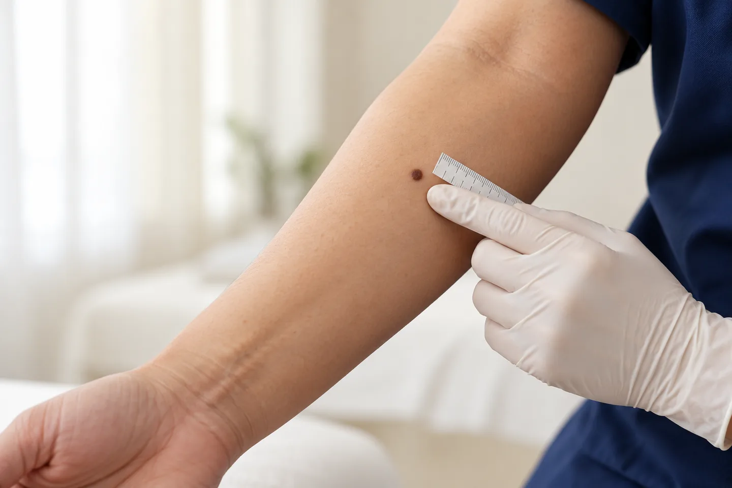
A mole check is a focused skin examination designed to assess whether a mole or pigmented lesion appears benign, needs monitoring, or should be biopsied to rule out melanoma or another skin cancer. Dermatologists commonly assess the lesion visually and, when appropriate, with dermoscopy, which magnifies structures and pigment patterns that cannot be fully appreciated with the naked eye.
For Thai patients, expats, tourists, and international patients in Bangkok, a mole check can be especially useful when there is a lesion that has recently changed, started bleeding, become itchy, developed irregular pigmentation, or simply looks different from the rest.
What Is a Mole?
A mole is a benign growth made up of pigment-producing cells called melanocytes.
Moles can be:
- Brown
- Tan
- Black
- Pink
- Skin-colored
- Blue-gray
- Flat
- Slightly raised
- Dome-shaped
A typical mole is often symmetrical, has relatively regular borders, and maintains a similar appearance over time. Moles may occur anywhere on the body, including the scalp, palms, soles, between the toes, and under or around the nails.
Having a mole does not mean it will become cancerous. Most remain benign throughout life.
Why Should a Mole Be Checked?
The main purpose of a mole check is to identify lesions that might represent melanoma or another form of skin cancer.
A mole deserves medical assessment particularly when it:
Appears recently and looks unusual
- Changes in size
- Changes in shape
- Changes in color
- Develops irregular borders
- Begins itching
- Becomes painful or tender
- Bleeds without obvious trauma
- Forms a crust repeatedly
- Develops a lump or thicker area
- Looks noticeably different from the patient’s other moles
A new or changing spot is one of the most important warning signs of melanoma.
What Is Melanoma?
Melanoma is a skin cancer that develops from melanocytes, the cells responsible for producing skin pigment.
It is less common than some other skin cancers but is clinically important because it can spread to other parts of the body if not diagnosed early.
Melanoma does not always begin in an existing mole. It may also develop as a completely new pigmented or non-pigmented lesion.
This is why mole surveillance should involve more than checking old moles alone.
How Can You Tell If a Mole Might Be Suspicious?
A useful screening guide is the ABCDE rule.
A — Asymmetry
One half of the lesion looks different from the other half.
A benign mole is often relatively symmetrical, although this rule is not absolute.
B — Border
Suspicious features include:
- Irregular edges
- Notched edges
- Blurred borders
- Poorly defined borders
C — Color
A mole may deserve assessment if it contains several colors or develops a new uneven color pattern.
Possible colors include:
- Light brown
- Dark brown
- Black
- Red
- Pink
- White
- Blue-gray
D — Diameter
A diameter greater than approximately 6 millimeters can be a warning feature.
However, melanoma can be smaller than 6 millimeters, so a small mole should not automatically be considered harmless.
E — Evolving
Change over time is particularly important.
This may include a change in:
- Size
- Shape
- Color
- Surface
- Thickness
- Symptoms
A changing lesion should be assessed even if it does not meet every other warning criterion.
The “Ugly Duckling” Sign
Many people have several moles that follow a similar pattern.
One mole that looks clearly different from all the others can be more significant than its size alone.
This is commonly called the ugly duckling sign.
For example, if almost all of a person’s moles are small and light brown but one lesion is much darker, irregular, or structurally different, that outlier may deserve closer examination.
This approach is particularly useful in people who have many moles.
Are All Irregular Moles Melanoma?
No.
Some benign moles can look unusual.
These are often called atypical moles.
An atypical mole may be:
- Larger than surrounding moles
- Irregularly shaped
- Unevenly colored
- Partly flat and partly raised
An atypical appearance does not automatically mean melanoma. However, people with multiple atypical moles may have a higher melanoma risk, and an individual lesion that changes or looks different from the person’s usual mole pattern deserves evaluation.
It is not generally recommended to remove every atypical mole simply because it looks unusual. Clinical assessment determines which lesions require monitoring or biopsy.
What Happens During a Mole Check?
A mole check generally begins with a medical history.
The dermatologist may ask:
- How long has the mole been present?
- Has it changed?
- Was the change sudden or gradual?
- Has it bled?
- Does it itch or hurt?
- Has it become darker or developed several colors?
- Have you had previous abnormal moles?
- Have you previously had melanoma or another skin cancer?
- Does anyone in your family have a history of melanoma?
- Do you have many moles?
- Have you had significant sun exposure or sunburns?
The lesion is then examined directly.
Depending on the clinical situation, the dermatologist may also examine surrounding skin or recommend a more complete skin examination.
What Is Dermoscopy?
Dermoscopy is a non-invasive examination using a handheld magnifying device designed for skin lesions.
It allows the dermatologist to see pigment structures, blood-vessel patterns, and other features beneath the surface of the skin that cannot be fully appreciated with the naked eye.
Dermoscopy can help distinguish many benign moles from lesions that require further investigation.
However, dermoscopy does not replace biopsy when a lesion remains suspicious.
Does a Suspicious Mole Always Need to Be Removed?
No.
After examination, several outcomes are possible.
The dermatologist may recommend:
- No treatment if the lesion appears clearly benign
- Self-monitoring
- Clinical photography
- Short-term review
- Digital dermoscopic monitoring
Biopsy or complete removal if melanoma cannot be confidently excluded
The appropriate approach depends on the lesion’s appearance, history, location, and the patient’s overall risk.
What Is Digital Mole Monitoring?
Some lesions look mildly atypical but do not have enough suspicious features to require immediate removal.
In selected cases, standardized photographs or digital dermoscopic images can be used to monitor whether the lesion changes over time.
Short-term digital monitoring can help distinguish stable benign lesions from lesions that evolve and therefore require biopsy.
Monitoring is only appropriate when the dermatologist considers it safe. A clearly suspicious lesion should not simply be photographed and left indefinitely.
When Is a Mole Biopsy Recommended?
A biopsy may be recommended when the dermatologist cannot confidently exclude melanoma or another skin cancer based on clinical and dermoscopic examination.
Reasons may include:
- Significant asymmetry
- Irregular structure
- Concerning pigment pattern
- Rapid change
- Unexplained bleeding
- New nodular growth
- Progressive enlargement
- A lesion that looks unlike all the patient’s other moles
Suspicious change on serial photographs
For a lesion suspicious for melanoma, complete removal for histopathological examination is generally preferred when technically appropriate because examining the whole lesion provides important diagnostic information.
What Happens to a Mole After It Is Removed?
The removed tissue can be sent to a pathology laboratory.
A pathologist examines the tissue under a microscope to determine what the lesion actually is.
Possible results can include:
- Benign mole
- Atypical or dysplastic mole
- Melanoma
- Another benign skin lesion
- Another type of skin cancer
The pathology result determines whether further treatment is necessary.
A mole should therefore not be destroyed cosmetically if there is meaningful concern about melanoma and tissue diagnosis is required.
Should Suspicious Moles Be Removed With Laser?
Laser treatment is generally not the preferred approach for a mole that is clinically suspicious for melanoma.
Destroying the pigment without obtaining adequate tissue may make pathological diagnosis impossible.
If there is concern about melanoma, obtaining tissue for histopathological examination is more important than cosmetic removal.
This distinction matters because harmless raised moles may sometimes be treated differently from suspicious pigmented lesions.
Can a Benign Mole Be Removed?
Yes.
A benign mole may sometimes be removed because it:
- Repeatedly catches on clothing
- Is irritated by jewelry
- Is cut while shaving
- Causes discomfort
- Is cosmetically unwanted
The method depends on whether the mole is raised or flat, its location, size, and whether tissue examination is needed.
A mole should first be assessed to determine that cosmetic removal is appropriate.
Can a Mole Grow Hair and Still Be Benign?
Yes.
Hair growing from a mole does not by itself mean the lesion is dangerous.
Benign moles can contain hair follicles.
What matters more is whether the lesion is changing in size, shape, color, structure, or symptoms.
Does Bleeding Mean a Mole Is Cancer?
Not always.
A raised benign mole can bleed after:
- Shaving
- Scratching
- Friction from clothing
- Accidental trauma
However, spontaneous or recurrent bleeding without a clear reason should be assessed, particularly when accompanied by other changes.
Is an Itchy Mole Dangerous?
Itching alone does not mean melanoma.
Many harmless skin lesions can itch because of dryness, irritation, or friction.
Nevertheless, a mole that develops new persistent itching together with visible change should be checked.
The same applies to new pain or tenderness.
Can Melanoma Be Smaller Than 6 mm?
Yes.
The diameter criterion in the ABCDE guide is useful, but melanoma can be diagnosed at a smaller size.
This is why change and overall appearance are more important than relying on size alone.
Can Melanoma Be Pink or Skin-Colored?
Yes.
Not every melanoma is dark brown or black.
Some melanomas contain relatively little pigment and can appear:
- Pink
- Red
- Skin-colored
Therefore, a new or changing bump should not be ignored merely because it lacks dark pigmentation.
Can Melanoma Develop on the Palms, Soles, or Nails?
Yes.
Melanoma can occur in areas that receive relatively little sun exposure, including:
- Palms
- Soles
- Between the toes
- Around the nails
- Under the nails
This is particularly important when examining patients with darker skin tones because melanoma in these patients may be more likely to occur in less sun-exposed areas such as the palms, soles, or nails.
A skin check should therefore not focus only on the face, arms, and back.
What Does Melanoma Under a Nail Look Like?
Possible signs include:
- A new brown or black vertical streak
- Progressive widening of the streak
- Irregular pigmentation
- Several shades of brown or black
- Pigment extending onto surrounding skin
- Nail splitting or destruction
- A lump underneath the nail
A dark streak does not automatically mean melanoma, especially in people with naturally pigmented nails, but a new or changing single-nail lesion deserves assessment.
What About Moles on the Scalp?
Moles and melanoma can occur on the scalp.
They may be difficult to notice because they are hidden by hair.
During self-examination, it can help to use:
- A mirror
- A hand mirror
- A family member or partner
- A hairdryer or comb to separate the hair
A dermatologist may also examine the scalp when performing a broader skin check.
What Is a Full-Body Mole Check?
A full-body mole check is different from looking at one specific mole.
It involves systematically examining the skin for:
- Suspicious moles
- New lesions
- Atypical moles
- Melanoma
- Other forms of skin cancer
Lesions that look different from the patient’s usual pattern
Depending on the reason for examination, areas checked may include:
- Scalp
- Face
- Ears
- Neck
- Chest
- Abdomen
- Back
- Arms
- Hands
- Legs
- Feet
- Between fingers and toes
- Palms
- Soles
- Nails
The exact extent of the examination should be discussed with the patient.
Who Should Consider a Full-Body Skin Check?
There is no universal schedule that is appropriate for everyone.
The need and frequency of professional skin examination should be individualized.
A dermatologist may recommend closer surveillance for people with factors such as:
- Previous melanoma
- Previous skin cancer
- Numerous moles
- Multiple atypical moles
- Strong family history of melanoma
- Significant ultraviolet exposure
- Repeated severe sunburns
- Immunosuppression
A concerning lesion that requires monitoring
Dermatologists advise people with many moles or increased melanoma risk to discuss the appropriate examination frequency with their dermatologist rather than assuming one schedule fits everyone.
How Often Should You Have Your Moles Checked?
There is no single correct interval for every person.
Some people may only need examination when they notice a new or changing lesion.
Others with higher melanoma risk may need scheduled surveillance.
The frequency depends on:
- Number of moles
- Presence of atypical moles
- Previous melanoma
- Previous skin cancer
- Family history
- Skin type
- Age
- Sun exposure
- Changes detected during previous examinations
A dermatologist can recommend an interval based on individual risk.
How to Check Your Moles at Home
Self-examination is useful because you are often the person most likely to notice that something has changed.
Try to examine the entire skin surface rather than only visible areas.
Useful equipment includes:
- A full-length mirror
- A hand mirror
- Good lighting
- Pay attention to:
- New spots
- Changing spots
- Moles that look different from the rest
- Non-healing lesions
- Bleeding lesions
- New lumps
Medical guidelines recommend checking areas that are easy to miss, including the scalp, soles, palms, nails, between the toes, and buttocks.
Should You Photograph Your Moles?
Photographs can be helpful for recognizing change.
When photographing a mole:
- Use similar lighting each time
- Keep the camera approximately the same distance away
- Include a wider image showing the location
- Take a close-up image
- Consider including a ruler for scale
However, photographs should not be used as a substitute for medical assessment when a lesion is already suspicious.
Can a Phone Application Diagnose a Melanoma?
A phone application should not be relied upon to diagnose or exclude melanoma.
Computer-assisted tools are developing rapidly, but concerns remain about diagnostic accuracy. Medical guidelines advise having concerning lesions assessed by a trained healthcare professional rather than relying solely on an application.
Does Bangkok’s Sun Increase Skin Cancer Risk?
Ultraviolet radiation contributes to the development of many skin cancers and is an important melanoma risk factor.
Bangkok and Thailand have substantial year-round ultraviolet exposure, making routine sun protection sensible for residents and visitors.
Useful measures include:
- Seeking shade during intense sun
- Wearing protective clothing
- Wearing a hat
- Using broad-spectrum sunscreen
- Reapplying sunscreen when appropriate
- Avoiding intentional tanning
However, not every melanoma develops because of recent sun exposure, and melanoma can occur on covered areas.
Are People With Asian Skin at Risk of Melanoma?
Yes.
Melanoma can occur in every skin tone.
Although the overall patterns of melanoma vary between populations, people with Asian or darker skin can develop melanoma, including on the palms, soles, and nails.
A common mistake is assuming that naturally darker skin eliminates melanoma risk.
It does not.
Any suspicious or changing lesion should be assessed regardless of skin color.
Should Every Mole Be Removed to Prevent Melanoma?
No.
Most moles are benign and do not need treatment.
Routine removal of every normal or atypical mole is not recommended.
Instead, the aim is to identify lesions that are genuinely suspicious or causing symptoms.
Even in people with atypical moles, melanoma can arise as a new lesion rather than necessarily developing from one of the existing moles.
When Should You See a Dermatologist Quickly?
Arrange assessment promptly if you notice:
- A new unusual mole
- A mole changing in size
- A mole changing in shape
- A mole changing in color
- A lesion becoming thicker
- Irregular borders
- Several colors within one lesion
- Unexplained bleeding
- Persistent crusting
- A new lump
- A lesion that does not heal
- A mole that looks very different from your other moles
- A new or changing dark streak under a nail
- Pigment spreading beyond a lesion
- A lesion that is rapidly evolving
These features do not automatically mean melanoma, but they justify professional assessment.
What to Expect During a Mole Check in Bangkok
A useful mole consultation should focus on diagnosis and risk assessment rather than immediately removing the lesion.
Before your appointment, consider:
- How long the mole has been present
- Whether it has changed
- When you first noticed the change
- Whether it bleeds or itches
- Previous skin cancers
- Previous abnormal mole biopsies
- Family history of melanoma
- Previous pathology reports
Photographs showing how the lesion looked several months or years earlier can be particularly useful.
During the consultation, the dermatologist may examine the lesion directly and use dermoscopy.
If the lesion looks reassuring, monitoring may be sufficient.
If it remains suspicious, biopsy or complete removal for pathological examination may be recommended.
For tourists and international patients in Bangkok, tell the dermatologist how long you will remain in Thailand because pathology results and further treatment may require follow-up.
Frequently asked questions
How do I know whether a mole is dangerous?
No single visual feature can diagnose melanoma.
Important warning signs include asymmetry, irregular borders, multiple colors, change over time, unexplained bleeding, and a lesion that looks different from your other moles.
Can a normal mole change slightly over time?
Yes. Some benign moles can slowly change over many years.
A rapid or noticeable recent change deserves more attention than a stable long-term appearance.
Is a large mole always dangerous?
No.
Size is only one feature.
A small changing lesion can be more concerning than a large mole that has remained stable for years.
Can melanoma be smaller than 6 mm?
Yes.
Melanoma can be smaller than 6 mm, so size alone cannot exclude it.
Does an irregular mole always mean melanoma?
No.
Atypical benign moles can have irregular shape or color.
Dermoscopy and clinical assessment help determine whether biopsy is necessary.
Should I worry if a mole is itchy?
New persistent itching should be assessed if it occurs together with visible change.
Itching alone does not necessarily mean cancer.
Should I worry if a mole bleeds?
Bleeding after shaving or obvious trauma may be harmless.
Repeated spontaneous bleeding deserves medical examination.
Can melanoma grow from an existing mole?
Yes, but melanoma can also develop as a completely new skin lesion.
This is why looking for both new and changing lesions is important.
What is dermoscopy?
Dermoscopy is a magnified examination of the skin that reveals structures not easily visible to the naked eye.
It helps dermatologists assess moles and other skin lesions.
Does dermoscopy hurt?
No.
It is a non-invasive examination and does not normally require anesthesia.
Can dermoscopy diagnose melanoma with certainty?
Not always.
If a lesion remains suspicious, pathological examination after biopsy or excision may still be necessary.
Does every unusual mole require a biopsy?
No.
Some can safely be monitored depending on their appearance and the patient's risk.
Should a suspicious mole be removed with laser?
Generally no if melanoma is a concern.
A suspicious lesion should usually be evaluated in a way that preserves tissue for pathological diagnosis.
Can benign moles be removed for cosmetic reasons?
Yes, after appropriate assessment.
The technique depends on the mole's appearance, depth, location, and whether tissue should be sent for pathology.
Do people with darker skin need mole checks?
Yes when clinically indicated.
Melanoma can occur in all skin tones and may involve the palms, soles, or nails in darker-skinned populations.
How often should I have a mole check?
The appropriate interval depends on individual risk.
People with previous melanoma, numerous atypical moles, or other significant risk factors may require more frequent surveillance.
Can I monitor a mole with photos?
Yes, photographs can help identify change, but a clearly suspicious lesion should still be examined by a dermatologist.
When should I see a dermatologist for a mole?
Arrange assessment if a mole is new, changing, bleeding, persistently itchy, structurally unusual, or clearly different from your other moles.
Related conditions
-
Full Body Skin Examination Full Body Skin Examination A full body skin examination is a head-to-toe assessment of the skin performed to look for suspicious moles, abnormal growths, precancerous changes and signs of skin cancer.

-
Melanoma Melanoma Melanoma is a serious type of skin cancer that develops from melanocytes, the cells that produce pigment in the skin. Unlike most basal cell carcinomas and many cutaneous squamous cell carcinomas, melanoma has a greater potential to spread to lymph nodes and other organs if it becomes invasive.

-
Dermoscopy Dermoscopy Dermoscopy is a non-invasive examination technique that allows a dermatologist to inspect skin lesions with greater detail than is possible with the naked eye alone.

-
Skin Biopsy (การวินิจฉัย / Diagnosis) Skin Biopsy (การวินิจฉัย / Diagnosis) A skin biopsy is a minor medical procedure in which a dermatologist removes a small sample of skin so it can be examined under a microscope. It is one of the most important diagnostic tools in dermatology because many skin conditions cannot be confirmed by appearance alone.