Full Body Skin Examination
A full body skin examination is a head-to-toe assessment of the skin performed to look for suspicious moles, abnormal growths, precancerous changes and signs of skin cancer.
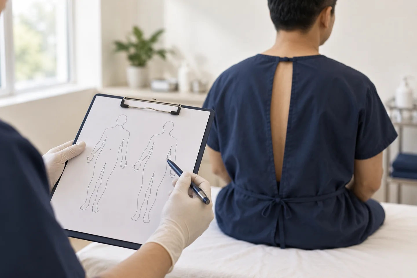
Unlike checking only one concerning mole, a complete examination evaluates areas that patients may not routinely see themselves, including the scalp, back, buttocks, backs of the legs, soles of the feet, between the toes and around the nails. Depending on the patient’s history and concerns, other less visible areas may also need examination.
During a full body examination, a dermatologist may use a handheld magnifying instrument called a dermatoscope to examine individual lesions more closely. If a lesion cannot be confidently identified as benign, the next step may be photography, monitoring or a skin biopsy depending on its appearance and clinical context. A biopsy is required to establish a definite diagnosis of skin cancer.
For patients living in Bangkok, expatriates, tourists and international patients, a skin examination can also provide an opportunity to review personal skin-cancer risk, previous sun exposure and which changes should be monitored at home.
What Is a Full Body Skin Examination?
A full body skin examination is a systematic visual examination of the skin from the scalp to the feet.
The purpose is not simply to count moles. A dermatologist looks for a broad range of abnormalities, including:
- Unusual or changing moles
- Possible melanoma
- Basal cell carcinoma
- Squamous cell carcinoma
- Precancerous sun damage
- Persistent non-healing lesions
- Unusual pigmented lesions
- New growths
- Suspicious nail changes
- Lesions that differ from a patient’s usual mole pattern
A full-body skin examination is a head-to-toe evaluation that includes the skin, scalp and nails.
Is a Full Body Skin Examination the Same as a Mole Check?
They overlap, but they are not exactly the same.
A mole check may focus mainly on melanocytic nevi and lesions that could represent melanoma.
A full body skin examination is broader. In addition to moles, the dermatologist may look for:
- Basal cell carcinoma
- Squamous cell carcinoma
- Actinic keratoses
- Unusual growths
- Persistent sores
- Pigmented lesions
- Nail abnormalities
- Other skin conditions discovered incidentally
If you have one particular mole that concerns you, tell the dermatologist before the examination so that it receives focused evaluation.
Why Have a Full Body Skin Examination?
One of the main reasons is that skin cancer does not always appear in an obvious or symptomatic way.
Some lesions:
- Do not hurt
- Do not itch
- Develop slowly
- Occur in areas that are difficult to see
- Look similar to an ordinary mole or age spot
- Appear in areas that receive little sun exposure
Melanoma can occur on sun-exposed skin, but it can also develop on the soles, beneath a nail or in other relatively protected areas. It can affect people of every skin color.
A professional skin examination therefore complements—not replaces—regular awareness of your own skin.
Who Should Consider a Full Body Skin Examination?
The appropriate frequency of professional skin examinations depends on individual risk. There is no single schedule that is appropriate for everyone.
A full body examination may be particularly useful for people with:
- A previous melanoma
- Previous basal cell carcinoma
- Previous squamous cell carcinoma
- Numerous moles
- Atypical-looking moles
- A strong family history of melanoma
- Significant lifetime ultraviolet exposure
- Frequent or severe sunburns
- Regular outdoor work or recreation
- Reduced immune function
- A lesion that has recently changed
- A new unexplained growth
- Difficulty examining their own skin
The interval between professional skin examinations should be individualized according to factors such as skin type, sun exposure, family history and previous skin cancer.
Do You Need a Skin Examination If You Have Darker Skin?
Potentially, yes.
Skin cancer is less common in some darker skin types, but it can still occur. Importantly, melanoma in darker skin can develop in areas such as:
- Soles of the feet
- Palms
- Beneath the nails
- Other less sun-exposed areas
Anyone, regardless of skin color, can develop melanoma.
This is particularly relevant in an international city such as Bangkok, where patients come from many ethnic and skin-type backgrounds. A proper examination should not be limited only to sun-exposed skin.
What Does a Dermatologist Look For During a Skin Examination?
The dermatologist is looking not only for a particular checklist of abnormal moles, but also for the overall pattern of your skin.
Important findings include:
- A new lesion
- A mole that has changed
- An unusual combination of colors
- Irregular shape
- Irregular borders
- A lesion unlike your other moles
- Persistent scaling
- Crusting
- Repeated bleeding
- A sore that does not heal
- A growth that repeatedly heals and returns
- A shiny or pearly bump
- A firm enlarging lesion
- Pigment under a nail
- An unexplained changing patch
One useful concept is the “ugly duckling” sign: a lesion that looks noticeably different from the patient’s other moles. A new or changing spot, or one that differs from surrounding lesions, can be an important melanoma warning sign.
The ABCDE Warning Signs of Melanoma
One widely used method for recognizing suspicious pigmented lesions is the ABCDE approach.
A — Asymmetry
One half of a mole does not resemble the other half.
B — Border
The edges may be irregular, blurred, scalloped or poorly defined.
C — Color
There may be several colors within one lesion rather than a uniform color.
These may include varying shades of:
- Brown
- Black
- Red
- Pink
- White
- Blue
D — Diameter
Larger lesions deserve attention, although melanoma can also be smaller than 6 millimeters.
E — Evolution
A lesion is changing in:
- Size
- Shape
- Color
- Surface
- Symptoms
Evolution is particularly important because change over time can be more meaningful than appearance alone.
The ABCDE system is useful, but not every melanoma follows all five rules. Any new or unusual changing lesion should be assessed if there is concern.
What Does Basal Cell Carcinoma Look Like?
Basal cell carcinoma can have several appearances.
Possible features include:
- A shiny or pearly bump
- A pink or reddish patch
- A sore that heals and then returns
- A lesion that bleeds easily
- A waxy or scar-like area
- A brown or darkly pigmented growth
Not every basal cell carcinoma looks the same, especially across different skin tones.
A lesion that repeatedly bleeds, crusts or fails to heal deserves evaluation.
What Does Squamous Cell Carcinoma Look Like?
Squamous cell carcinoma can appear as:
- A rough or scaly patch
- A crusted bump
- A persistent sore
- A firm enlarging growth
- A lesion that bleeds
- A rough red patch
- A thickened area
Squamous cell carcinoma may take several forms, which is another reason that self-diagnosis from photographs alone can be unreliable.
Does a Skin Examination Hurt?
No. The examination itself is visual and should not be painful.
The dermatologist may:
- Inspect your skin
- Move hair to examine the scalp
- Look between fingers and toes
- Examine palms and soles
- Inspect nails
- Use a dermatoscope
Nothing is removed during the examination unless you and the physician decide that a biopsy or another procedure is appropriate.
What Is Dermoscopy?
Dermoscopy is examination of a skin lesion using a handheld instrument called a dermatoscope.
It combines magnification with specialized illumination, allowing the dermatologist to see colors and structures beneath the skin surface that are not as clearly visible to the naked eye.
Dermatoscopy can help distinguish among:
- Benign moles
- Seborrheic keratoses
- Vascular lesions
- Some basal cell carcinomas
- Some squamous lesions
- Suspicious melanocytic lesions
Dermoscopy can be used as an effective tool during professional mole and skin examination.
Dermoscopy improves clinical assessment, but it does not replace biopsy when tissue diagnosis is necessary.
Does Every Mole Need Dermoscopy?
Not necessarily.
An experienced dermatologist may recognize many common benign lesions clinically.
Dermoscopy is especially useful for:
- Pigmented lesions
- Atypical moles
- Lesions that do not have a clear clinical diagnosis
- Comparing similar-looking lesions
- Evaluating structures that cannot be seen clearly without magnification
The dermatologist decides which lesions require closer examination.
What Is Mole Mapping?
Mole mapping generally refers to systematic photographic documentation of moles over time.
Depending on the method, it can involve:
- Standard body photographs
- Close-up lesion photography
- Dermoscopic images
- Digital comparison at future visits
The goal is not simply to produce photographs. The value lies in comparing lesions over time and identifying:
- New moles
- Changing lesions
- Subtle structural changes
Mole mapping can be particularly useful for selected higher-risk patients with many moles or atypical mole patterns. Photographic monitoring programs can be helpful in people at higher risk of melanoma.
It is not automatically necessary for every patient.
Full Body Skin Examination vs Mole Mapping
These are complementary rather than interchangeable.
| Full Body Skin Examination | Mole Mapping |
|---|---|
| Dermatologist examines the skin directly | Photographs document lesions |
| Can assess non-pigmented skin cancers | Most useful for monitoring moles |
| Provides assessment at that visit | Helps compare change over time |
| Dermoscopy may be used | Dermoscopic photography may be included |
| Can identify lesions requiring biopsy | Helps identify new or changing lesions |
Dermatologist examines the skin directly Photographs document lesions Can assess non-pigmented skin cancers Most useful for monitoring moles Provides assessment at that visit Helps compare change over time Dermoscopy may be used Dermoscopic photography may be included Can identify lesions requiring biopsy Helps identify new or changing lesions A patient may need one, both or neither depending on their individual risk.
What Areas Are Included in a Full Body Skin Examination?
A thorough skin examination may include:
- Scalp
- Face
- Ears
- Neck
- Chest
- Abdomen
- Back
- Arms
- Underarms
- Hands
- Fingernails
- Buttocks
- Legs
- Feet
- Soles
- Between the toes
- Toenails
Depending on history, risk factors and symptoms, examination of other covered areas may also be appropriate.
For example, after a melanoma diagnosis, a complete examination includes checking the scalp, skin, nails and, when clinically indicated, less visible areas such as genital skin and the mouth.
Patient comfort, privacy and consent should always be respected.
Do I Need to Remove All My Clothes?
For a true full-body skin examination, most skin needs to be visible.
Patients are generally asked to undress to an appropriate level and may be provided with a gown or covering.
The examination should be performed in a private setting. Areas can be uncovered sequentially rather than exposing the entire body at once.
If there is an area you do not want examined, tell the dermatologist. Examination should always proceed with consent.
Full skin checks generally require removal of most clothing, with a gown used for modesty where appropriate.
Should the Scalp Be Checked?
Yes.
The scalp is easy to miss during self-examination because hair can hide lesions.
The dermatologist may separate the hair section by section to inspect the scalp.
Patients should mention:
- A scalp growth
- A recurring scab
- A bleeding spot
- A lesion noticed by a hairdresser
- A new pigmented area
If you have photographs of a scalp lesion taken previously, they may be useful for comparison.
Should the Feet and Soles Be Checked?
Yes.
Melanoma can occur on the soles, including in people with darker skin.
A full examination may therefore include:
- Tops of the feet
- Soles
- Heels
- Between the toes
- Toenails
Suspicious pigment beneath a nail or on a sole should not automatically be assumed to be trauma or bruising if it persists or changes.
Are Nails Checked During a Full Body Skin Examination?
They may be.
A dermatologist may look for:
- Pigmented streaks
- New dark areas
- Pigment spreading onto surrounding skin
- Persistent nail destruction
- Unexplained abnormalities
Many nail changes are benign and are not melanoma. However, persistent unexplained changes may need further evaluation.
What Happens If the Dermatologist Finds a Suspicious Mole?
Finding a suspicious lesion does not automatically mean that it is cancer.
Depending on the lesion, the dermatologist may recommend:
- No treatment
- Routine observation
- Clinical photography
- Dermoscopic photography
- Short-term follow-up
- Skin biopsy
The decision depends on the lesion’s appearance, history and level of suspicion.
What Is a Skin Biopsy?
A skin biopsy removes part or all of a lesion so it can be examined under a microscope by a pathologist.
A biopsy may be recommended if the dermatologist suspects:
- Melanoma
- Basal cell carcinoma
- Squamous cell carcinoma
- Another tumor
- A skin condition that cannot be diagnosed confidently from examination alone
A skin biopsy is required for definitive diagnosis when skin cancer is suspected.
Does a Suspicious Mole Always Need to Be Removed?
No.
Many unusual-looking lesions are benign.
The decision to remove or biopsy a lesion depends on factors such as:
- Clinical appearance
- Dermoscopic findings
- Whether it is new
- Whether it has changed
- Symptoms
- Patient age
- Location
- Personal history
- Previous skin cancer
- Whether reliable monitoring is possible
Removing every mole is neither necessary nor desirable.
Can a Dermatologist Tell Whether a Mole Is Cancer Just by Looking?
Sometimes a lesion may look very strongly suspicious or clearly benign, but visual examination alone cannot provide absolute certainty in every case.
If melanoma or another skin cancer is suspected, pathological examination of tissue is the definitive diagnostic step.
This is why statements such as “this is definitely not cancer” should be made cautiously when a lesion has uncertain clinical features.
How Often Should You Have a Full Body Skin Examination?
There is no universal interval that applies to everyone.
Frequency should depend on individual risk.
Someone with no significant risk factors may require a different schedule from someone with:
- Previous melanoma
- Previous non-melanoma skin cancer
- Numerous atypical moles
- Strong family history
- Significant ultraviolet exposure
- Reduced immune function
Individualized examination frequency is recommended based on risk factors and previous skin cancer history.
Patients who have previously had melanoma generally require structured long-term dermatologic follow-up rather than relying on general population screening schedules.
Is an Annual Skin Examination Necessary for Everyone?
Not necessarily.
A yearly examination may be appropriate for some people, particularly those at increased risk, but there is no single evidence-based annual schedule that must apply to every asymptomatic person.
A more clinically sensible approach is to personalize follow-up according to:
- Age
- Skin type
- Number of moles
- Previous skin cancers
- Family history
- Sun exposure
- Previous biopsies
- Immune status
- Findings on the initial examination
Patients should also seek evaluation between scheduled appointments if they notice a new or changing lesion.
Can a Full Body Skin Examination Detect Every Skin Cancer?
No medical screening examination is perfect.
A full-body examination can identify many suspicious lesions, but limitations include:
- Very early lesions may be subtle
- A lesion may develop after the examination
- Some areas may be difficult to visualize
- Certain melanomas do not follow classic appearance rules
- Some lesions require biopsy for diagnosis
- Changes over time may be more informative than a single examination
That is why professional examinations and patient self-awareness work best together.
How Should I Prepare for a Full Body Skin Examination?
Preparation is simple.
Before your appointment:
- Avoid heavy makeup if facial lesions need evaluation
- Remove nail polish if possible when nail examination is important
- Avoid covering concerning lesions
- Bring previous pathology reports if available
- Bring photographs of changing lesions
- Bring previous mole-mapping images if you have them
- Make a list of lesions you specifically want checked
- Know whether close relatives have had melanoma or other skin cancers
If you have had previous biopsies abroad, pathology reports can be especially useful for a dermatologist in Bangkok.
Should I Point Out a Mole Even During a Full Body Examination?
Yes.
Do not assume the doctor automatically knows which lesion worries you.
Tell the dermatologist if a lesion:
- Recently appeared
- Changed
- Bleeds
- Itches persistently
- Hurts
- Keeps crusting
- Heals and returns
- Looks different from your other moles
A patient’s observation of change over time is valuable because the dermatologist is seeing the lesion at only one moment.
How Long Does a Full Body Skin Examination Take?
The duration varies considerably depending on:
- Number of moles
- Number of suspicious lesions
- Whether dermoscopy is needed
- Whether photographs are taken
- Previous skin cancer history
- Whether biopsies or treatment discussions are required
A focused screening can be relatively quick, whereas examination of a patient with hundreds of moles or complex previous melanoma history may take considerably longer.
The quality and thoroughness of the examination matter more than a fixed number of minutes.
What Happens After the Examination?
Possible outcomes include:
Everything appears benign
The dermatologist may recommend:
- Routine self-monitoring
- Sun protection
- Follow-up based on individual risk
A lesion is worth monitoring
The lesion may be:
- Measured
- Photographed
- Examined dermoscopically
- Rechecked after a defined interval
A lesion requires biopsy
The dermatologist may recommend biopsy for pathological diagnosis.
A benign lesion is found
Examples may include:
- Seborrheic keratoses
- Cherry angiomas
- Skin tags
- Benign moles
These do not necessarily require removal unless there is a diagnostic question, irritation or another appropriate reason.
Can Benign Moles Become Cancer?
Most ordinary benign moles never become melanoma.
Melanoma can develop from a pre-existing mole, but it can also appear as a completely new lesion.
Therefore, the goal is not to remove every mole. Instead, patients should monitor for unusual change and have suspicious lesions evaluated.
Should Every Mole Be Removed to Prevent Melanoma?
No.
Routine removal of all normal moles is not an appropriate melanoma-prevention strategy.
Instead, prevention and early detection focus on:
- Recognizing suspicious change
- Appropriate professional evaluation
- Sun protection
- Monitoring high-risk patients
- Biopsy of clinically concerning lesions
What Is the “Ugly Duckling” Sign?
Most people’s moles share a relatively consistent pattern.
A lesion that looks very different from the others may deserve closer attention.
This is known as the ugly duckling sign.
For example, if most of your moles are:
- Small
- Light brown
- Round
- Uniform
but one lesion is:
- Black
- Irregular
- Larger
- Multicolored
that difference may be clinically meaningful.
A spot that looks different from the rest is an important melanoma warning sign.
Can Skin Cancer Be Colorless?
Yes.
Not every melanoma or skin cancer is dark brown or black.
Some skin cancers may appear:
- Pink
- Red
- Flesh-colored
- Pearly
- White
- Scar-like
- Crusted
Therefore, checking only dark moles can miss other forms of skin cancer.
When Should a Spot Be Checked Quickly?
Arrange medical assessment if you notice:
- A rapidly changing mole
- A new unusual pigmented lesion
- A mole changing color
- New asymmetry
- Irregular borders
- A lesion that looks different from all others
- Unexplained bleeding
- Persistent crusting
- A non-healing sore
- A lesion that heals and returns
- Rapid growth
- Persistent unexplained pigment beneath a nail
Prompt medical evaluation is recommended for new spots or lesions changing in size, shape or color.
Is It Normal for a Mole to Itch?
Temporary itching does not automatically indicate cancer.
Moles can itch because of:
- Dry skin
- Friction
- Irritation
- Eczema
- Trauma
However, persistent itching associated with change, bleeding, enlargement or altered appearance should be evaluated.
What About a Mole That Bleeds?
Bleeding may occur from trauma, such as shaving or scratching.
However, spontaneous or repeated bleeding without obvious injury deserves medical assessment, particularly if the lesion is changing.
What About a Sore That Will Not Heal?
A persistent sore is an important reason to seek evaluation.
Basal cell carcinoma and squamous cell carcinoma can sometimes appear as a sore that:
- Does not heal
- Crusts repeatedly
- Bleeds
- Appears to heal and then returns
Such a lesion should not simply be treated repeatedly as irritation without reassessment.
Skin Self-Examination Between Dermatology Visits
A professional examination does not replace checking your own skin.
Many clinicians recommend regular self-examination, often approximately monthly, particularly for higher-risk individuals.
A practical self-check should include:
- Face
- Ears
- Neck
- Chest
- Abdomen
- Arms
- Underarms
- Hands
- Nails
- Back
- Buttocks
- Legs
- Feet
- Soles
- Between the toes
- Scalp
Use a full-length mirror and handheld mirror, and consider help from a partner for difficult-to-see areas.
What Should You Look for During a Self-Examination?
Look for:
- New spots
- Changing moles
- Unusual colors
- Irregular borders
- Enlarging lesions
- Persistent sores
- Recurrent crusting
- Bleeding lesions
- A spot that looks unlike your others
Knowing your own baseline skin pattern makes it easier to recognize change.
Should You Take Photographs of Your Moles?
Photographs can be useful when used sensibly.
They may help document:
- Size
- Shape
- Location
- Change over time
However, photographs should not be used to reassure yourself about a lesion that is clearly changing or concerning.
Image quality, lighting and camera angle can also make comparison unreliable.
If you cannot obtain prompt medical assessment, serial photographs may provide useful information for the eventual consultation, but they are not a substitute for clinical examination.
Are Skin Cancer Phone Applications Reliable?
Applications that attempt to classify moles from photographs are increasingly available, but they should not replace dermatologist evaluation.
There are continuing concerns regarding the accuracy of artificial-intelligence-based skin-cancer applications, and more evidence is needed before they can be relied upon as a substitute for professional evaluation.
Sun Exposure and Skin Cancer Risk in Thailand
Living in Thailand can involve substantial ultraviolet exposure throughout the year.
Patients may accumulate exposure through:
- Outdoor commuting
- Beach activities
- Running or cycling
- Golf
- Swimming
- Outdoor work
- Travel within tropical regions
Sun protection remains relevant even when a full skin examination is normal.
General measures include:
- Seeking shade
- Wearing protective clothing
- Wearing a broad-brimmed hat
- Using appropriate sunscreen on exposed skin
- Avoiding intentional tanning
- Reapplying sunscreen when needed
Broad-spectrum, water-resistant sunscreen with a sun protection factor of at least 30 is recommended as part of ultraviolet protection.
Does Having a Tan Protect Against Skin Cancer?
A tan should not be considered reliable protection from ultraviolet damage.
Intentional ultraviolet tanning adds to cumulative ultraviolet exposure and is not recommended as a skin-cancer prevention strategy.
Full Body Skin Examination for Expats in Bangkok
Expatriates may have accumulated substantial sun exposure before moving to Thailand, particularly if they previously lived in:
- Australia
- New Zealand
- Europe
- North America
- South Africa
- Other high-ultraviolet environments
Risk assessment should therefore consider lifetime exposure, not simply how long someone has lived in Bangkok.
Patients who have had previous biopsies or skin cancers in another country should bring:
- Pathology reports
- Surgical reports
- Previous skin photographs
- Mole-mapping records
- Previous dermatologist letters
Full Body Skin Examination for Tourists and International Patients
A visitor may seek a skin examination in Bangkok because:
- A mole changed during travel
- A partner noticed a lesion
- A lesion has started bleeding
- A previously monitored mole needs review
They want a routine examination while in Thailand
If a suspicious lesion is identified and follow-up will occur in another country, obtaining a clear written diagnosis, photographs where appropriate and copies of pathology reports can help maintain continuity of care.
What to Expect During a Dermatology Consultation in Bangkok
Before the examination, the dermatologist may ask about:
- Previous melanoma
- Previous basal cell carcinoma
- Previous squamous cell carcinoma
- Previous atypical moles
- Previous biopsies
- Family history of melanoma
- Number of moles
- Sunburn history
- Outdoor work
- Tanning-bed exposure
- Immune suppression
- Current medications
- A specific lesion that concerns you
The examination may then proceed systematically from head to toe.
A dermatoscope may be used for selected lesions.
At the end of the examination, the dermatologist should explain which lesions appear benign, whether any lesions require monitoring and whether biopsy is recommended.
Questions to Ask During Your Skin Examination
Useful questions include:
- Are any of my moles atypical?
- Is there one lesion I should monitor especially closely?
- Do I have an increased melanoma risk?
- How often should I check my own skin?
- How often should I have a professional skin examination?
- Do I need photographic monitoring?
- Does this lesion need biopsy or can it be observed?
- What specific change should prompt an earlier appointment?
The goal is to leave knowing not only what the examination found, but also what you should monitor going forward.
Frequently asked questions
What is a full body skin examination?
It is a systematic head-to-toe examination of the skin to look for suspicious moles, skin cancers, precancerous lesions and other abnormal growths.
Is a full body skin examination the same as skin cancer screening?
The terms are often used interchangeably, although a complete dermatology examination may evaluate more than skin cancer alone.
Who should have a full body skin examination?
It can be particularly useful for people with previous skin cancer, many moles, atypical moles, strong family history, significant ultraviolet exposure or a concerning changing lesion.
Do I need an annual skin check?
Not everyone needs the same schedule. Examination frequency should be based on individual risk factors and previous skin-cancer history.
What does a dermatologist check during a skin examination?
A dermatologist may examine the scalp, face, ears, neck, trunk, arms, hands, nails, legs, feet, soles and other areas as clinically appropriate.
Do I have to undress?
A true full-body examination requires most skin to be visible. A gown or covering can be used, and examination should respect privacy and consent.
Does a skin examination hurt?
No. It is primarily visual.
What is dermoscopy?
Dermoscopy uses a magnifying instrument with specialized lighting to examine structures within a skin lesion more closely.
Can dermoscopy diagnose melanoma without biopsy?
Dermoscopy helps determine whether a lesion is suspicious, but definitive diagnosis of suspected melanoma requires pathological examination of tissue.
What is mole mapping?
Mole mapping uses systematic photography to document lesions so that new or changing moles can be recognized over time.
Does everyone need mole mapping?
No. It is generally most useful for selected patients at increased melanoma risk or those with many moles.
What is the difference between a mole check and mole mapping?
A mole check evaluates lesions during the appointment. Mole mapping creates photographic records for comparison over time.
What does melanoma look like?
Melanoma may appear as a new or changing lesion, an irregular mole, a multicolored lesion or a spot that looks different from the patient's other moles.
What does the ABCDE rule mean?
It refers to asymmetry, irregular border, uneven color, diameter and evolution or change.
Can melanoma be smaller than 6 millimeters?
Yes. Diameter is only one clue; smaller lesions can still be melanoma.
Can melanoma be pink or colorless?
Yes. Not every melanoma is darkly pigmented.
Can melanoma occur on the feet?
Yes. Melanoma can develop on the soles and other relatively sun-protected areas.
Can people with dark skin develop melanoma?
Yes. Melanoma can occur in every skin color.
Should nails be examined for melanoma?
Suspicious persistent pigmentation or changing nail abnormalities may require examination.
Does every unusual mole need biopsy?
No. Some can be confidently identified as benign or monitored, while suspicious lesions may require biopsy.
Does biopsy mean the doctor thinks I definitely have cancer?
No. Biopsy is performed to establish the diagnosis when clinical examination alone cannot provide sufficient certainty.
Can a dermatologist tell whether a mole is cancer by looking at it?
Some lesions can be strongly suspected clinically, but definitive diagnosis of skin cancer requires biopsy and microscopic examination.
Should all moles be removed?
No. Most benign moles do not require removal.
Can normal moles turn into melanoma?
Some melanomas arise within existing moles, but many develop as new lesions.
What is the ugly duckling sign?
It describes a lesion that looks noticeably different from the patient's other moles and therefore may deserve closer assessment.
Should I worry if a mole itches?
Temporary itching has many benign causes. Persistent itching accompanied by change, bleeding or growth deserves assessment.
Should a bleeding mole be checked?
Yes, particularly when bleeding is recurrent or occurs without obvious trauma.
What should I do about a sore that does not heal?
A persistent non-healing or repeatedly crusting lesion should be examined because some skin cancers can appear this way.
How often should I check my own skin?
Many clinicians recommend regular self-examination, commonly around once a month, with frequency adjusted for individual risk.
Can I photograph my moles at home?
Yes, photographs can help document changes, but they should not replace medical evaluation of a suspicious lesion.
Can a mobile application tell me whether a mole is cancer?
Such applications have limitations and should not replace evaluation by a trained clinician.
How should I prepare for a full body skin examination?
Bring previous pathology reports or photographs, identify lesions you are worried about and remove nail polish if nail examination is relevant.
Should I bring old biopsy reports?
Yes, particularly if you have previously had melanoma, atypical moles or other skin cancers.
Can I have a skin examination while visiting Bangkok?
Yes. For international patients, previous dermatology records can be particularly helpful for comparison and continuity of care.
When should I see a dermatologist urgently rather than wait for a routine skin check?
Seek earlier assessment for rapidly changing lesions, recurrent unexplained bleeding, rapidly growing growths or persistent non-healing sores.
Related conditions
-
Mole Check Mole Check Most moles are harmless. However, a new mole, a changing mole, or a spot that looks different from your other moles deserves closer attention.
-
Melanoma Melanoma Melanoma is a serious type of skin cancer that develops from melanocytes, the cells that produce pigment in the skin. Unlike most basal cell carcinomas and many cutaneous squamous cell carcinomas, melanoma has a greater potential to spread to lymph nodes and other organs if it becomes invasive.

-
Basal Cell Carcinoma Basal Cell Carcinoma Basal cell carcinoma (BCC) is a common type of skin cancer that usually grows slowly and is most often found on sun-exposed skin. Although BCC rarely spreads to distant parts of the body, it can continue growing into surrounding skin and deeper structures if left untreated. Treatment is therefore recommended after the diagnosis is confirmed.

-
Squamous Cell Carcinoma Squamous Cell Carcinoma Squamous cell carcinoma, often abbreviated as SCC, is a common form of skin cancer arising from squamous cells in the epidermis, the outer layer of the skin. It frequently develops on chronically sun-exposed areas such as the face, scalp, ears, lips, forearms and backs of the hands, although it can occur elsewhere.