





























Squamous Cell Carcinoma
Squamous cell carcinoma, often abbreviated as SCC, is a common form of skin cancer arising from squamous cells in the epidermis, the outer layer of the skin. It frequently develops on chronically sun-exposed areas such as the face, scalp, ears, lips, forearms and backs of the hands, although it can occur elsewhere.
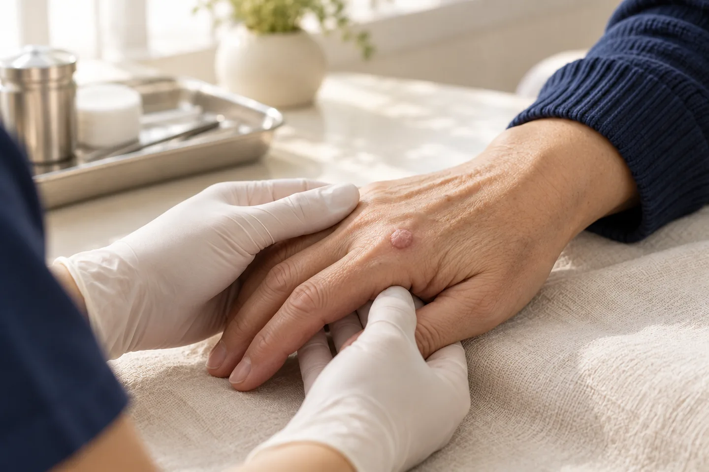
Most localized squamous cell carcinomas can be treated effectively, and surgery is the main treatment for many patients. However, SCC deserves careful assessment because some tumors can grow deeply, invade nearby structures, involve nerves, spread to lymph nodes or, less commonly, spread to distant organs. The treatment plan therefore depends not only on whether SCC is present, but also on whether the tumor has low-risk or high-risk features. Surgical excision, Mohs micrographic surgery, radiation therapy and selected nonsurgical approaches all have roles in appropriate patients.
For Thai patients, expatriates, tourists and international patients seeking skin cancer treatment in Bangkok, the most useful first step is usually to establish the diagnosis with an appropriate examination and biopsy, review the pathology carefully, and then select treatment according to the individual tumor rather than using the same procedure for every SCC.
What Is Squamous Cell Carcinoma?
Squamous cell carcinoma of the skin is a malignant tumor arising from keratinocytes, the cells that make up much of the epidermis.
It belongs to the group of keratinocyte skin cancers, together with basal cell carcinoma. Although many SCCs remain localized and can be treated with surgery, SCC generally has a greater potential for deeper invasion and metastatic spread than typical basal cell carcinoma.
Treatment approaches for localized cutaneous SCC include surgery, Mohs micrographic surgery, radiation therapy, curettage and electrodesiccation, and cryosurgery. More advanced disease may require systemic immunotherapy.
Squamous Cell Carcinoma vs Basal Cell Carcinoma
Both are common skin cancers, but their behavior is different.
Basal cell carcinoma usually grows locally and only very rarely spreads to distant sites.
Squamous cell carcinoma can also be slow-growing, but some tumors behave more aggressively. High-risk SCC can invade deeper tissue, grow along nerves or spread to regional lymph nodes.
This difference is why determining the risk category of an SCC is important after diagnosis.
What Causes Squamous Cell Carcinoma?
Long-term ultraviolet exposure is one of the major causes of cutaneous SCC.
Risk accumulates over many years. A patient’s history of sun exposure before moving to Thailand may be just as relevant as their current exposure in Bangkok.
Important risk factors include:
- Cumulative ultraviolet exposure
- Repeated sunburn
- Outdoor work or recreation
- Increasing age
- Previous SCC or other skin cancer
- Fair skin that burns easily
- Reduced immune function
- Organ transplantation
- Immunosuppressive medication
- Previous radiation treatment
- Certain chronic wounds, scars or inflammatory skin conditions
- Certain genetic disorders associated with increased skin-cancer risk
Living in a sunny climate such as Thailand makes ongoing ultraviolet protection particularly important, but SCC can occur in all skin tones.
Can People With Darker or Asian Skin Develop SCC?
Yes.
Darker pigmentation lowers some ultraviolet-related skin cancer risks but does not eliminate skin cancer.
SCC in darker skin can also arise in areas not typically associated with heavy sun exposure, including chronic scars, wounds and inflammatory skin conditions. Patients should therefore not dismiss a persistent ulcer, enlarging growth or changing lesion simply because they have darker skin.
What Does Squamous Cell Carcinoma Look Like?
SCC does not have one single appearance.
It may present as:
- A firm pink, red or skin-colored bump
- A rough or thickened scaly patch
- A crusted growth
- A dome-shaped nodule
- A sore that does not heal
- A lesion that repeatedly bleeds or crusts
- A rapidly enlarging lump
- A tender or painful growth
- A wart-like lesion
- An ulcerated lesion
Some lesions resemble benign conditions such as warts, seborrheic keratoses, inflamed cysts or chronic dermatitis. This is one reason suspicious lesions should be evaluated medically rather than destroyed without diagnosis.
Warning Signs That Should Be Evaluated
A dermatologist should assess a lesion if it:
- Does not heal
- Continues to enlarge
- Bleeds repeatedly
- Develops persistent crusting
- Becomes painful or unusually tender
- Becomes thick or firm
- Grows rapidly
- Appears in a chronic scar or wound
- Returns after previous treatment
- Develops numbness, tingling or unusual nerve-like pain
- Is accompanied by an enlarging nearby lymph node
Rapid growth, significant pain, neurological symptoms or an enlarged lymph node deserve particular attention because they may indicate a higher-risk tumor.
What Is Actinic Keratosis?
Actinic keratosis is a sun-damaged precancerous skin lesion.
It often appears as a persistent rough, scaly or gritty patch on sun-exposed skin, particularly on the face, bald scalp, ears, forearms and hands.
Actinic keratosis is not invasive SCC, but some lesions can progress to SCC. Treatment is therefore often recommended depending on the clinical situation. The National Cancer Institute distinguishes actinic keratosis from skin cancer but recognizes it as a lesion treated because of its potential to develop into cancer.
What Is Squamous Cell Carcinoma in Situ?
Squamous cell carcinoma in situ means abnormal cancerous squamous cells are confined to the epidermis and have not invaded deeper skin.
It is also commonly known as Bowen disease.
It may appear as a:
- Persistent red patch
- Scaly plaque
- Crusted area
- Slowly enlarging patch
- Lesion resembling eczema or psoriasis
Because it has not invaded beyond the epidermis, SCC in situ is biologically different from invasive SCC and may have a broader range of treatment options.
How Is Squamous Cell Carcinoma Diagnosed?
Diagnosis usually starts with a dermatological examination.
The dermatologist evaluates:
- Size
- Location
- Border
- Surface
- Thickness
- Ulceration
- Growth rate
- Bleeding
- Pain or tenderness
- Previous treatment
- Previous skin-cancer history
A dermatoscope may also be used to examine structures within the lesion that cannot easily be seen with the naked eye.
However, when SCC is suspected, skin biopsy and histopathological examination are generally required to confirm the diagnosis and provide information needed for treatment planning.
What Type of Biopsy Is Used?
The biopsy technique depends on the lesion.
Possible approaches include:
Shave Biopsy
A superficial portion of the lesion is removed.
Punch Biopsy
A small cylindrical sample containing deeper skin layers is taken.
Incisional Biopsy
Only part of a larger lesion is removed for diagnosis.
Excisional Biopsy
The entire visible lesion is removed when appropriate.
For suspected SCC, the biopsy should obtain enough tissue to establish the diagnosis and, where possible, provide information about invasion and other clinically important features.
What Does the Pathology Report Tell Us?
The pathology report can provide much more than the words “squamous cell carcinoma.
”
Important information may include:
- Whether SCC is present
- Whether it is in situ or invasive
- Degree of differentiation
- Tumor thickness or depth when assessable
- Presence of perineural invasion
- Other aggressive histological features
- Margin status after excision
These findings are interpreted together with the tumor’s size, location, clinical appearance and patient risk factors.
What Does Well-Differentiated or Poorly Differentiated SCC Mean?
Differentiation describes how closely the cancer cells resemble normal squamous cells under the microscope.
A well-differentiated SCC retains more features of normal squamous cells.
A poorly differentiated SCC looks more abnormal microscopically and can be associated with more aggressive behavior.
Differentiation is only one component of risk assessment and should not be interpreted in isolation.
What Is Perineural Invasion?
Perineural invasion means cancer cells are found growing around or along a nerve.
This is an important pathological finding because it can be associated with a higher-risk tumor and may influence treatment planning.
Depending on the situation, additional surgery, imaging, radiation treatment or multidisciplinary assessment may be considered.
Symptoms such as persistent pain, numbness, tingling, weakness or unusual sensation around an SCC should therefore be mentioned to the treating doctor.
Low-Risk vs High-Risk Squamous Cell Carcinoma
Not all SCCs behave the same way.
Risk assessment is central to treatment planning.
Risk stratification is recommended for localized cutaneous SCC because tumor characteristics influence both treatment selection and prognosis.
Factors that may increase concern include:
- High-risk anatomical location
- Larger tumor size
- Greater depth
- Poorly defined borders
- Rapid growth
- Poor differentiation
- Perineural invasion
- Tumor recurrence
- Previous radiation at the site
- Immunosuppression
- Certain aggressive pathological characteristics
A small well-defined SCC on a lower-risk site may therefore be treated differently from a recurrent poorly differentiated lesion on the ear or scalp.
Why Does Tumor Location Matter?
Certain anatomical sites require special consideration.
Examples include:
- Ear
- Lip
- Eyelid
- Nose
- Scalp
- Hands
- Feet
- Genital skin
These locations may be associated with higher-risk behavior, greater functional importance, or both.
Treatment must therefore balance adequate cancer removal with preservation of normal tissue and function.
Can SCC Spread to Lymph Nodes?
Yes.
Most localized SCCs have not spread when diagnosed, but cutaneous SCC can metastasize, most commonly first to nearby regional lymph nodes.
Risk is higher in certain aggressive tumors.
For higher-risk SCC, the dermatologist or treating specialist may therefore examine regional lymph nodes as part of the consultation.
Does Every SCC Patient Need Scans?
No.
Small, localized, lower-risk SCC generally does not require routine imaging.
Imaging may be considered when there is concern about:
- Deep invasion
- Bone involvement
- Perineural spread
- Regional lymph-node disease
- Recurrent high-risk tumor
- Very large tumor
- Other clinical features suggesting advanced disease
The appropriate study depends on what the treating team needs to evaluate.
Squamous Cell Carcinoma Treatment Options
For localized SCC, treatment options include:
- Standard surgical excision
- Mohs micrographic surgery
- Radiation therapy
- Curettage and electrodesiccation in selected low-risk lesions
- Cryosurgery in selected situations
For advanced SCC that cannot be treated adequately with local therapy, systemic immunotherapy may be considered.
Surgical Excision for Squamous Cell Carcinoma
Standard surgical excision is widely used for SCC.
The surgeon removes:
the visible tumor + an appropriate margin of surrounding apparently normal skin
The tissue is then sent for histopathological examination.
Surgery is generally considered the most effective treatment for cutaneous SCC.
Advantages of Surgical Excision
Excision:
- Removes the tumor
- Provides tissue for pathology
- Allows margin assessment
- Can be used for many localized SCCs
- Can often be completed in one procedure
Limitations
Possible disadvantages include:
- Scar formation
- Bleeding
- Bruising
- Infection
- Wound separation
- Temporary or persistent numbness
- Need for a skin flap or graft in larger defects
- Need for further surgery if margins are involved
How Wide Should the Surgical Margin Be?
There is no single margin that is correct for every SCC.
The appropriate margin depends on tumor characteristics such as:
- Diameter
- Anatomical site
- Border definition
- Histological differentiation
- Tumor risk category
- Previous treatment
Professional references describe wider margins for larger, less differentiated or higher-risk tumors than for small lower-risk lesions. However, surgical margins should be individualized rather than selected from one universal number.
What Does a Clear Surgical Margin Mean?
After excision, the pathologist examines sections from the specimen margins.
A pathology report may describe the margins as:
- Clear
- Negative
- Free of tumor
This generally means cancer cells were not identified at the examined tissue edge.
Margin status is important, but it must be interpreted together with the type of surgery and the tumor’s risk characteristics.
What Does a Positive Margin Mean?
A positive or involved margin means SCC reaches the examined edge of the specimen.
This raises concern that microscopic tumor may still remain.
Depending on the tumor, further management may include:
- Additional excision
- Mohs micrographic surgery
- Radiation therapy in selected situations
- Other individualized treatment
A high-risk SCC with involved margins deserves especially careful management.
What Is Mohs Surgery?
Mohs micrographic surgery is a specialized form of skin cancer surgery in which the tumor is removed in stages.
After the visible tumor and a thin layer of surrounding tissue are removed, the margins are examined microscopically.
If cancer remains in a particular area, the surgeon removes another layer specifically from that location.
This continues until no cancer is identified at the examined margins.
The technique allows detailed margin assessment while conserving uninvolved tissue.
When Is Mohs Surgery Considered for SCC?
Mohs surgery can be particularly useful for SCC that:
- Develops on an ear, eyelid, scalp or hand
- Occurs near the eye or ear
- Has poorly defined borders
- Has returned after previous treatment
- Has an increased risk of recurrence or spread
- Appears to involve a nerve or blood vessel
- Develops in previously irradiated skin
These are examples rather than absolute rules. The final decision depends on the individual tumor and patient.
Mohs Surgery vs Standard Excision
| Standard Excision | Mohs Surgery |
|---|---|
| Tumor removed with a planned surrounding margin | Tumor removed in mapped stages |
| Tissue usually processed through routine pathology | Margins examined during the procedure |
| Appropriate for many localized SCCs | Particularly useful for selected high-risk SCCs |
| Further surgery may be needed if margins are involved | Residual cancer can be targeted during the same procedure |
| Often straightforward | Offers detailed margin control and tissue conservation |
Neither procedure is automatically better for every patient.
The best choice depends on the cancer.
Does Every Facial SCC Need Mohs Surgery?
No.
A facial location alone does not automatically mean that every SCC requires Mohs surgery.
The dermatologist considers:
- Exact anatomical site
- Tumor size
- Histological subtype
- Border definition
- Whether it is recurrent
- Depth
- Other high-risk features
Mohs can be particularly valuable in anatomically important or high-risk areas, but standard excision may remain appropriate for selected facial tumors.
Curettage and Electrodesiccation
This technique involves scraping tumor tissue with a curette and treating the base with electrical energy.
It may be considered for carefully selected low-risk SCCs.
Its main limitation is that it does not provide the same type of comprehensive surgical margin assessment as excision or Mohs surgery.
It is therefore not generally appropriate for tumors with significant high-risk features.
Can SCC Be Frozen?
Cryosurgery destroys tissue by freezing it.
It may have a role in selected low-risk cases or when surgery is unsuitable.
Its limitations include:
- Lack of conventional margin assessment
- Uncertainty about the exact depth of destruction
- Potential pigment change or scarring
- Limited suitability for high-risk tumors
Nonsurgical treatments such as cryosurgery may be considered for selected low-risk tumors or when surgery is contraindicated, although outcomes may be less favorable than surgical treatment.
Can Laser Treat Squamous Cell Carcinoma?
Laser destruction is not a standard first-line treatment for invasive cutaneous SCC.
This distinction is important because patients may mistake SCC for a wart, seborrheic keratosis or another benign growth.
Destroying a suspicious lesion before obtaining adequate diagnostic tissue may make it harder to determine:
- Whether it was cancer
- How deeply it invaded
- Whether aggressive features were present
- Whether the cancer was completely removed
Suspicious lesions should therefore be diagnosed before cosmetic destruction.
Can SCC Be Treated With Cream?
This depends heavily on whether the lesion is SCC in situ or invasive SCC.
Topical treatments may sometimes be considered for selected SCC in situ lesions.
They are not routine substitutes for surgical management of invasive SCC.
If a lesion is thick, rapidly growing, ulcerated or otherwise suspicious for invasion, it should be appropriately assessed and biopsied before topical treatment is chosen.
Photodynamic Therapy
Photodynamic therapy may have a role in selected superficial or in-situ disease.
Photodynamic therapy is a possible treatment for SCC in situ rather than a standard treatment for invasive SCC.
Its suitability depends on the lesion, location, pathology and patient.
Radiation Therapy for SCC
Radiation therapy uses high-energy radiation to damage cancer cells.
It may be considered when:
- Surgery is unsuitable
- Surgery would cause substantial functional or anatomical difficulty
- A tumor is in a challenging location
- Disease has recurred
- Additional treatment is required after surgery in selected higher-risk situations
Radiation therapy is an established treatment option for localized SCC and can also be incorporated into management of recurrent or advanced disease.
Limitations of Radiation Therapy
Potential considerations include:
- Multiple treatment visits
- Temporary skin inflammation
- Long-term pigment or vascular changes
- Fibrosis or texture changes
- No surgical specimen confirming complete removal
- Difficulty re-treating the same site in some situations
It is therefore selected according to the individual patient’s circumstances.
What Is Advanced Squamous Cell Carcinoma?
Advanced cutaneous SCC can refer to disease that:
- Invades deeply into underlying structures
- Involves nerves, muscle or bone
- Spreads to regional lymph nodes
- Spreads to distant organs
- Cannot reasonably be treated with curative local surgery or radiation alone
These cases require a different pathway from a small localized skin cancer.
Management may involve dermatology, surgical oncology, radiation oncology, medical oncology, head-and-neck surgery or other specialties depending on the tumor.
Immunotherapy for Advanced SCC
Some locally advanced or metastatic SCCs can be treated with immune checkpoint inhibitors.
These medicines help the immune system recognize and attack cancer cells.
PD-1 inhibitors such as cemiplimab and pembrolizumab are used for selected advanced cutaneous SCC.
These medicines are not treatments for routine small localized SCC.
They are systemic cancer therapies and may cause immune-related adverse effects involving organs such as the skin, bowel, liver, lungs, endocrine glands and others. Treatment therefore requires appropriate oncology supervision.
Can SCC Come Back After Treatment?
Yes.
No treatment can guarantee that SCC will never recur.
Recurrence risk depends on factors including:
- Tumor location
- Tumor size
- Depth
- Histological features
- Perineural invasion
- Immune status
- Previous treatment
- Margin status
- Whether the tumor had already recurred
Recurrent nonmetastatic SCC is generally considered higher risk and may be treated with additional excision, Mohs surgery or radiation depending on the situation.
What Does Recurrent SCC Look Like?
Possible warning signs around a previous treatment site include:
- A new lump
- Persistent crusting
- Recurrent ulceration
- Bleeding
- Progressive thickening
- Pain
- Numbness
- A changing surgical scar
Any persistent change within or next to a previous SCC scar should be evaluated rather than automatically attributed to scar tissue.
Can SCC Spread Along Nerves?
Yes.
Perineural spread is an important feature of certain high-risk SCCs.
Patients should mention symptoms such as:
- Persistent localized pain
- Numbness
- Tingling
- Burning
- Weakness
- Changes in sensation
These symptoms do not prove nerve involvement, but they may influence the investigation and treatment plan.
What Happens if a Lymph Node Is Enlarged?
An enlarged lymph node does not automatically mean metastatic cancer.
Lymph nodes can enlarge due to infection and inflammation.
However, a persistent or suspicious lymph node near a known high-risk SCC should be assessed.
Depending on the situation, evaluation may involve imaging and tissue sampling.
Follow-Up After Squamous Cell Carcinoma
Follow-up serves two purposes:
Detecting recurrence of the treated SCC.
Detecting additional new skin cancers.
Ongoing skin examinations are recommended after SCC, with at least annual assessment and frequency adjusted according to individual risk.
Higher-risk patients may require closer surveillance.
Should I Have a Full-Body Skin Examination After SCC?
Often, yes.
A patient who has developed one SCC is also at increased risk of developing additional skin cancers.
Follow-up should therefore not focus only on the surgical scar.
A broader examination may identify:
- Additional SCCs
- Basal cell carcinoma
- Actinic keratoses
- Suspicious pigmented lesions
- Melanoma
Recovery After SCC Surgery
Recovery depends on:
- Tumor size
- Anatomical location
- Type of surgery
- Size of the surgical defect
- Method of wound closure
- Patient health
- Medications affecting bleeding or healing
- Temporary symptoms can include:
- Soreness
- Swelling
- Bruising
- Mild bleeding
- Tightness
- Numbness near the incision
How Is the Wound Closed?
After the tumor is removed, the wound may be managed with:
- Direct stitches
- Healing naturally without stitches
- Local skin flap
- Skin graft
- More complex reconstruction when necessary
A larger defect after surgery does not necessarily mean that anything went wrong. Skin cancers can extend microscopically beyond the visible surface.
The priority is adequate cancer treatment before cosmetic reconstruction.
Will SCC Surgery Leave a Scar?
Yes.
Any procedure that cuts through the skin leaves some degree of scar.
The eventual scar depends on:
- Tumor size
- Location
- Depth
- Closure method
- Skin tension
- Individual healing
- Infection or other complications
Scars generally change for months after surgery and often become less prominent as they mature.
Possible Complications of SCC Surgery
Potential complications include:
- Bleeding
- Infection
- Bruising
- Swelling
- Wound separation
- Delayed wound healing
- Scarring
- Pigment change
- Numbness
- Altered sensation
- Need for further surgery
- Tumor recurrence
The likelihood and significance of each complication vary with the individual operation.
Wound Care After Surgery
Follow the wound-care instructions provided by the treating clinician.
General principles may include:
- Keeping the wound clean
- Using recommended dressings
- Avoiding unnecessary trauma
- Temporarily limiting activities that stretch the wound
- Attending wound reviews
- Having sutures removed when required
- Seek medical assessment if there is:
- Persistent significant bleeding
- Increasing pain
- Progressive redness or swelling
- Pus
- Fever associated with worsening wound symptoms
- Wound separation
Can I Fly After SCC Surgery?
Many patients seeking dermatological care in Bangkok are tourists or international patients.
Whether flying is appropriate depends on:
- Size and location of surgery
- Type of reconstruction
- Bleeding risk
- Dressing requirements
- Suture removal schedule
- Timing of the final pathology report
- Whether additional surgery might be required
If you have upcoming international travel, discuss it before surgery rather than after the procedure has already been performed.
Can I Have SCC Treated in Bangkok and Follow Up Overseas?
Potentially, yes.
Continuity of care is especially important for skin cancer.
Before travelling, patients should ideally have access to:
- Pathology report
- Procedure or operative report
- Margin status
- Wound-care instructions
- Suture-removal plan
- Recommendations for additional treatment
- Follow-up recommendations
For higher-risk tumors, arranging appropriate follow-up before leaving Thailand is particularly important.
What Should International Patients Bring to a Consultation?
Bring any available:
- Pathology report
- Previous biopsy report
- Surgical report
- Clinical photographs
- Dermoscopic photographs
- Imaging reports
- Medication list
- Previous skin-cancer history
For complex cases, original pathology slides or tissue blocks may sometimes be useful for specialist review.
What to Expect During an SCC Consultation in Bangkok
A consultation may include several components.
Examination of the Tumor
The dermatologist evaluates its size, location, thickness, border and surface.
Review of Symptoms
Particular attention may be paid to:
- Rapid growth
- Pain
- Bleeding
- Numbness
- Tingling
- Previous recurrence
Skin Cancer History
Previous SCC, basal cell carcinoma, melanoma and actinic keratoses are relevant.
Medication and Immune Status
Immunosuppressive medications and organ transplantation can significantly influence risk.
Lymph Node Examination
For higher-risk tumors, nearby regional lymph nodes may be examined.
Pathology Review
If biopsy has already been performed, the pathology result is reviewed together with the clinical findings.
Treatment Planning
The discussion may include:
- Standard excision
- Mohs surgery
- Radiation therapy
- Additional investigation
- Referral for multidisciplinary treatment when necessary
When Should You See a Dermatologist?
Arrange an assessment if you have:
- A non-healing sore
- A rapidly growing skin lesion
- A painful or tender growth
- Persistent bleeding or crusting
- A firm scaly lump
- A lesion on the ear or lip
- A suspicious lesion developing in a chronic wound or scar
- Recurrence at a previous skin-cancer site
- Seek prompt review if a known or suspected SCC is associated with:
- Rapid enlargement
- Significant pain
- Numbness or neurological symptoms
- A growing regional lymph node
- Deep fixation to underlying structures
Frequently asked questions
Is squamous cell carcinoma dangerous?
Many localized SCCs are treatable, but some tumors can invade deeply or spread to lymph nodes and other sites. Risk assessment is therefore important.
Is SCC more dangerous than basal cell carcinoma?
In general, cutaneous SCC has a greater metastatic potential than typical basal cell carcinoma.
Can squamous cell carcinoma spread?
Yes. Spread can occur, particularly in higher-risk SCC.
Where does SCC usually spread first?
Regional lymph nodes are an important potential site of metastasis.
What does SCC look like?
It can appear as a firm bump, rough scaly patch, crusted growth, ulcer or non-healing sore.
Can SCC look like a wart?
Yes. Some SCCs have a wart-like or keratotic surface.
Does SCC hurt?
Some SCCs are painless; others may be tender, painful or associated with abnormal sensation.
Does SCC grow quickly?
Some grow slowly, while others can enlarge rapidly. Rapid growth should be evaluated.
Does SCC bleed?
It can, particularly if ulcerated or traumatized.
How is SCC diagnosed?
Diagnosis is generally confirmed with a skin biopsy and histopathological examination.
Can dermoscopy diagnose SCC?
Dermoscopy can support clinical assessment, but it does not replace histopathology when cancer needs confirmation.
What is SCC in situ?
It is SCC confined to the epidermis without deeper invasion. It is also known as Bowen disease.
Is actinic keratosis the same as SCC?
No. Actinic keratosis is a precancerous sun-damaged lesion, although some lesions can progress to SCC.
What is the best treatment for SCC?
There is no single best treatment for every SCC. Surgery is the principal treatment for many localized tumors, with the choice of technique based on risk.
Does SCC always need surgery?
Not every SCC is treated identically, but surgery is the most effective and commonly used treatment for invasive localized SCC.
What is Mohs surgery?
Mohs surgery removes skin cancer in stages while examining the margins microscopically during the procedure.
Does every facial SCC need Mohs surgery?
No. The decision depends on exact location, size, pathology, recurrence and other risk factors.
When is Mohs surgery recommended?
It is particularly useful for selected high-risk, recurrent, poorly defined or anatomically important tumors.
What does a clear margin mean?
It means cancer was not seen at the examined edge of the surgical specimen.
What if the margin is positive?
Further treatment may be required because residual tumor may remain.
Can SCC be treated with cream?
Selected SCC in situ lesions may sometimes be treated topically. Topical therapy is not routinely used as a substitute for surgery in invasive SCC.
Can SCC be frozen?
Cryosurgery may be considered for selected lower-risk lesions, but it has important limitations.
Can SCC be treated with laser?
Laser destruction is not a standard first-line treatment for invasive SCC.
Can radiation treat SCC?
Yes. Radiation therapy can be used in selected localized, recurrent or higher-risk cases.
What is high-risk SCC?
High-risk SCC has clinical or pathological characteristics associated with increased risk of recurrence, invasion or metastasis.
What is perineural invasion?
It means cancer is growing around or along a nerve and is an important high-risk feature.
Does every SCC patient need a scan?
No. Imaging is generally reserved for selected higher-risk or clinically concerning cases.
Do lymph nodes need to be checked?
For higher-risk SCC, examination of regional lymph nodes may be appropriate.
What happens if SCC spreads?
Treatment may involve surgery, radiation, immunotherapy or a combination of approaches.
What is immunotherapy for SCC?
Immunotherapy stimulates or releases restraints on the immune response against cancer. PD-1 inhibitors such as cemiplimab and pembrolizumab are used in selected advanced SCC.
Can SCC come back after treatment?
Yes. Recurrence can occur, especially in tumors with higher-risk features.
Can SCC recur even with clear margins?
It is possible. Margin status is important but does not eliminate all future recurrence risk.
What does recurrent SCC look like?
It may appear as a new lump, ulcer, crusting, pain, numbness or progressive change around a previous scar.
Should I have my whole skin checked after SCC?
Often yes, because patients who develop one SCC have an increased risk of additional skin cancers.
How often should I have a skin check?
Follow-up should be individualized according to tumor and patient risk, with at least annual skin examinations recommended after SCC.
Will SCC surgery leave a scar?
Yes. Any surgical procedure through the skin creates a scar.
How long does SCC surgery take to heal?
Healing time varies significantly according to tumor location, defect size and reconstruction.
Can I exercise after SCC surgery?
Activity restrictions depend on the location and method of wound closure.
Can I fly after SCC surgery?
Often possible, but plans should consider wound care, bleeding risk, sutures, pathology results and possible need for additional treatment.
Can tourists have SCC treated in Bangkok?
Potentially, provided enough time is available for diagnosis, pathology, treatment and follow-up.
Can pathology from another country be reviewed in Thailand?
Previous pathology reports are useful. In selected cases, review of original pathology slides or tissue may also be appropriate.
Related conditions
-
Basal Cell Carcinoma Basal Cell Carcinoma Basal cell carcinoma (BCC) is a common type of skin cancer that usually grows slowly and is most often found on sun-exposed skin. Although BCC rarely spreads to distant parts of the body, it can continue growing into surrounding skin and deeper structures if left untreated. Treatment is therefore recommended after the diagnosis is confirmed.

-
Melanoma Melanoma Melanoma is a serious type of skin cancer that develops from melanocytes, the cells that produce pigment in the skin. Unlike most basal cell carcinomas and many cutaneous squamous cell carcinomas, melanoma has a greater potential to spread to lymph nodes and other organs if it becomes invasive.

-
Bowen’s Disease Bowen's Disease Bowen’s disease, also called squamous cell carcinoma in situ, is an early form of squamous cell skin cancer in which abnormal cancerous cells remain confined to the outermost layer of the skin, the epidermis. Because the abnormal cells have not invaded into the deeper skin, Bowen’s disease is considered non-invasive. It is nevertheless a true form of skin cancer and should usually be treated or monitored because a small proportion of lesions can progress to invasive squamous cell carcinoma.

-
Actinic Keratosis / Precancerous Lesions Actinic Keratosis / Precancerous Lesions A rough, scaly spot that repeatedly crusts, feels tender, or does not heal may be more than simple dry or sun-damaged skin. Actinic keratosis (AK) is a precancerous skin lesion caused mainly by cumulative ultraviolet (UV) damage and is particularly important to recognize because some lesions can progress to cutaneous squamous cell carcinoma (SCC).
