Photodamage
Photodamage is the cumulative injury caused by repeated exposure to ultraviolet radiation from sunlight and artificial UV sources. Over time, it can lead to uneven pigmentation, sun spots, fine lines, rough texture, visible blood vessels, loss of elasticity and, importantly, precancerous or cancerous skin changes.
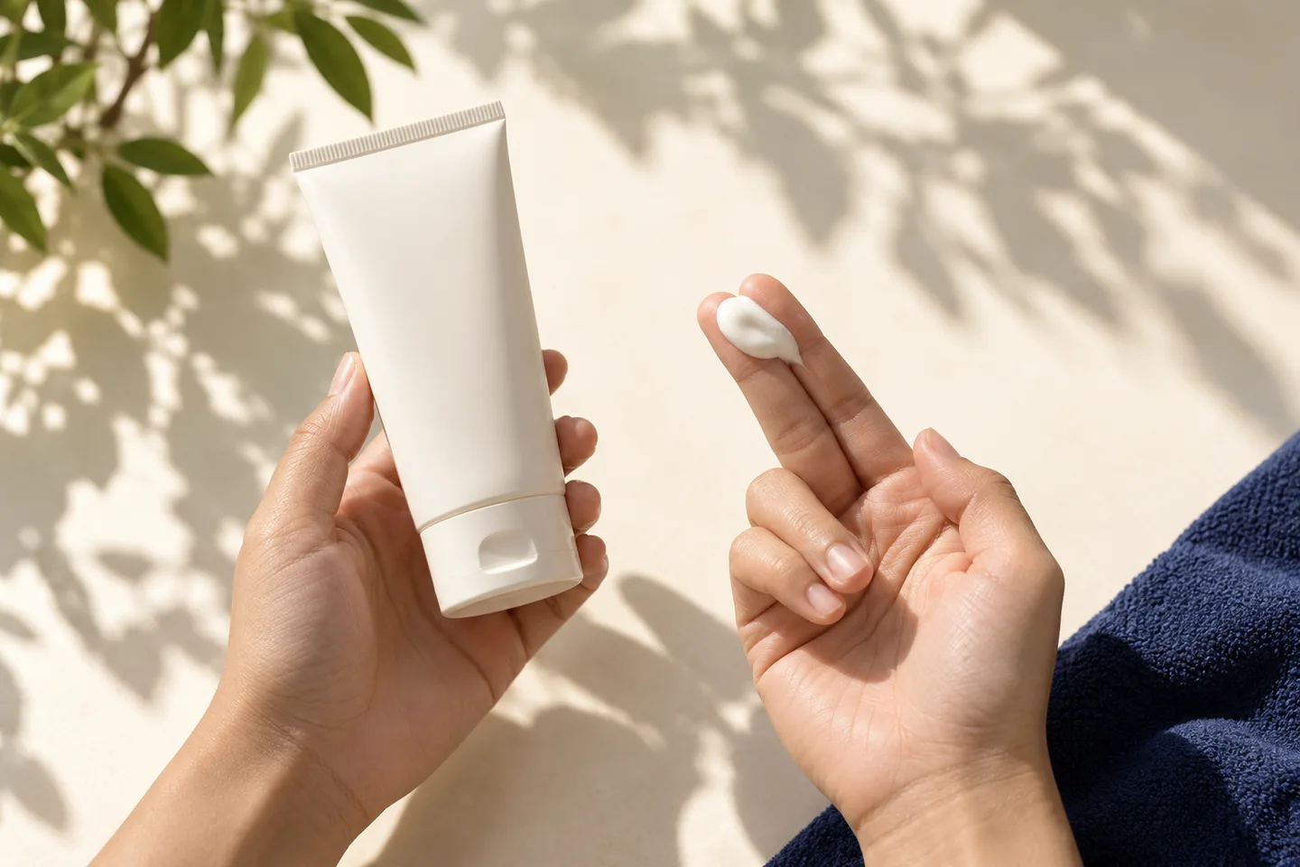
Not every brown spot, rough patch or wrinkle on sun-exposed skin is simply cosmetic. Some changes are benign, such as solar lentigines, while others may represent actinic keratoses or skin cancer and should be medically assessed before cosmetic treatment.
For patients in Bangkok and Thailand, where UV exposure can be significant throughout the year, effective management of photodamage usually involves two separate goals: preventing further UV injury and treating the specific changes that have already developed.
What Is Photodamage?
Photodamage refers to skin changes caused by cumulative ultraviolet exposure.
The term photoaging is often used when discussing the visible aging effects of sun exposure, such as wrinkles, pigmentation and loss of skin elasticity.
Natural aging and photoaging occur together, but they are not exactly the same.
Natural aging happens gradually because of biological changes within the skin and underlying tissues.
Photodamage is caused primarily by external ultraviolet exposure and can accelerate or exaggerate many signs normally associated with aging.
Wrinkles, age spots, loose skin and uneven or ruddy pigmentation are among the common visible manifestations of photoaging. Chronic sun damage can also lead to actinic keratoses and skin cancer.
What Causes Photodamage?
The main cause is repeated exposure to ultraviolet radiation.
Ultraviolet A and Ultraviolet B
Both UVA and UVB contribute to skin damage, although they affect the skin in somewhat different ways.
Repeated ultraviolet exposure can damage:
- DNA within skin cells
- collagen
- elastin
- pigment-producing pathways
- blood vessels
- other components of the skin’s supporting structure
The cumulative effect becomes increasingly visible over time.
Who Is Most at Risk of Photodamage?
Anyone exposed to ultraviolet radiation can develop photodamage, but the degree varies considerably.
Important risk factors include:
- frequent outdoor activity
- repeated sunburns
- long-term residence in sunny or tropical climates
- outdoor occupations
- insufficient sun protection
- use of tanning beds or artificial ultraviolet sources
- lighter skin that burns easily
- increasing age
- immunosuppression
- previous actinic keratoses or skin cancer
People with darker skin have more natural protection against some ultraviolet effects but are not immune to photodamage or skin cancer.
The appearance of photodamage may also differ according to skin tone.
Why Photodamage Is Relevant in Bangkok and Thailand
Thailand has a tropical climate with substantial potential for ultraviolet exposure throughout much of the year.
For people living in Bangkok, cumulative exposure can come not only from deliberate sunbathing but from routine activities such as:
- commuting
- walking outdoors
- outdoor exercise
- driving
- eating or working outdoors
- beach and island travel
- golf, tennis and other outdoor sports
Tourists and international patients may also experience intense intermittent exposure, especially when combining city travel with beach destinations.
Photodamage therefore should not be thought of only as a problem affecting people who intentionally sunbathe.
What Does Photodamaged Skin Look Like?
Photodamage can affect pigmentation, texture, blood vessels and skin structure simultaneously.
Common features include:
Uneven Pigmentation
The skin may develop areas of irregular brown pigmentation, mottling or uneven tone.
Several different conditions can produce brown spots, so not every area of pigmentation should automatically be treated as a cosmetic sun spot.
Solar Lentigines
Solar lentigines are flat brown patches caused by chronic ultraviolet exposure.
They are commonly called:
- sun spots
- age spots
- liver spots
Despite the name “liver spot,
” they are unrelated to liver disease.
Solar lentigines are benign and do not require treatment unless the patient wishes to improve their appearance. However, some skin cancers and precancerous lesions can resemble solar lentigines.
A dermatologist may therefore recommend examination before laser or other pigment-removing treatment if a lesion is atypical.
Fine Lines and Wrinkles
Long-term ultraviolet exposure damages structural components of the skin, including collagen.
This contributes to:
- fine lines
- deeper wrinkles
- reduced elasticity
- thinner or more fragile skin
- uneven surface texture
Photodamage-related wrinkles frequently occur together with natural age-related facial changes.
Rough or Uneven Skin Texture
Photodamaged skin may become:
- dry
- coarse
- rough
- uneven
- less elastic
Some areas may feel rough before they become particularly noticeable visually.
A persistent rough or scaly spot on a sun-exposed area deserves attention because it may represent an actinic keratosis rather than ordinary dryness.
Visible Blood Vessels and Redness
Chronic ultraviolet exposure can contribute to dilated superficial blood vessels, particularly on the face.
Patients may notice:
- persistent redness
- small visible vessels
- blotchy pigmentation mixed with redness
These vascular changes may coexist with other conditions such as rosacea, so diagnosis is important before treatment.
Actinic Keratosis: When Sun Damage Becomes Precancerous
One of the most important distinctions in any discussion of photodamage is the difference between cosmetic photoaging and actinic keratosis.
Actinic keratosis is a precancerous lesion caused by ultraviolet-related damage to skin cells. It often occurs on chronically sun-exposed areas such as:
- face
- ears
- nose
- forehead
- bald scalp
- backs of the hands
- forearms
It may appear as a rough, scaly or crusted area and can be easier to feel than to see.
Actinic keratoses are considered precancerous because some can progress to cutaneous squamous cell carcinoma.
This is why a persistent rough spot should not simply be covered with skincare products or treated cosmetically without evaluation.
Warning Signs That Need Dermatologic Evaluation
Photodamaged skin may contain both harmless and medically important lesions.
See a dermatologist if you notice a lesion that:
- continues to grow
- becomes thicker
- bleeds repeatedly
- develops a persistent crust
- ulcerates
- becomes painful or tender
- changes shape or colour
- develops irregular pigmentation
- looks different from your other spots
- repeatedly heals and returns
- does not heal
- changes noticeably over a short period
Tender, thickened, ulcerated or enlarging actinic keratoses are particularly concerning for possible progression toward squamous cell carcinoma and warrant medical assessment.
A suspicious lesion should generally be diagnosed before cosmetic laser treatment.
Can Photodamage Cause Skin Cancer?
Yes.
Ultraviolet radiation is an important cause of several forms of skin cancer and precancerous skin disease.
Chronic photodamage may be associated with:
- actinic keratosis
- squamous cell carcinoma
- basal cell carcinoma
- melanoma
This does not mean that every person with sun spots or wrinkles has skin cancer.
Most visible changes of photoaging are benign.
However, extensive photodamage indicates substantial cumulative ultraviolet exposure, which is one reason regular skin assessment can be useful in appropriate patients.
How Dermatologists Diagnose Photodamage
Diagnosis begins with a clinical examination.
A dermatologist may evaluate:
- distribution of pigmentation
- colour and shape of brown spots
- skin texture
- fine and deeper wrinkles
- elasticity
- redness and visible vessels
- actinic keratoses
- suspicious moles or lesions
- evidence of previous sun damage
Depending on the findings, additional assessment may include Dermoscopy.
Dermoscopy
Dermoscopy uses a magnifying device with specialized lighting to examine structures within pigmented and non-pigmented skin lesions.
It can help differentiate among conditions such as:
- solar lentigo
- seborrheic keratosis
- actinic keratosis
- melanocytic lesions
- certain forms of skin cancer
Dermoscopy is particularly helpful when a lesion looks similar to a simple sun spot but has atypical features.
When Is a Skin Biopsy Needed?
A biopsy is not necessary for ordinary cosmetic photodamage.
However, it may be recommended when a lesion is clinically suspicious or when the diagnosis remains uncertain.
For example, a dermatologist may consider biopsy if a lesion raises concern for:
- squamous cell carcinoma
- basal cell carcinoma
- melanoma
- atypical actinic keratosis
- another unusual skin growth
A biopsy involves removing a small sample of skin for examination under a microscope.
If a lesion is suspicious, diagnosis takes priority over cosmetic treatment.
Treatment of Photodamage
There is no single treatment that corrects every component of photodamage.
The best approach depends on what the patient actually has.
A person whose main problem is brown sun spots requires a different treatment from someone whose main concern is rough actinic keratoses, facial redness, wrinkles or skin laxity.
Treatment may therefore involve several approaches.
1. Sun Protection: The Foundation of Treatment
No photodamage treatment plan is complete without reducing further ultraviolet exposure.
Sun protection helps prevent additional damage and can reduce the development of further sun-related skin changes.
Useful measures include:
- broad-spectrum sunscreen
- appropriate protective clothing
- hats
- sunglasses
- shade
- reducing unnecessary exposure during intense sunlight
Patients should reapply sunscreen when appropriate, especially during prolonged outdoor activity, sweating or swimming.
Sun protection is particularly important after procedures that temporarily make the skin more sensitive to light.
2. Topical Retinoids
Topical Retinoids are vitamin A derivatives used in dermatology for acne and photoaged skin.
Prescription Retinoids may improve selected signs of photodamage, including:
- fine wrinkles
- rough texture
- irregular pigmentation
They work gradually and require consistent use.
Potential side effects include:
- dryness
- peeling
- redness
- irritation
- increased sensitivity
Treatment usually needs to be introduced gradually.
Patients with eczema, Rosacea or sensitive skin may require a modified regimen.
Pregnancy or pregnancy planning should be discussed with a doctor before using topical Retinoids.
3. Treatments for Solar Lentigines and Sun Spots
When a brown lesion has been confirmed to be benign, several treatments may be considered for cosmetic improvement.
Options can include:
- pigment-targeting Laser
- Intense Pulsed Light
- Cryotherapy
- Chemical peel
- selected topical medications
Solar lentigines are benign and do not require medical treatment unless desired for cosmetic reasons. Treatment can improve them, but recurrence or development of new lesions remains possible because ultraviolet exposure continues over time.
4. Pigment-Targeting Laser
Certain lasers selectively target melanin within pigmented lesions.
They may be useful for appropriately diagnosed solar lentigines and other selected pigmentation concerns.
Advantages
- treatment can target individual lesions
- often useful for discrete sun spots
- surrounding skin can often be relatively preserved
Limitations
Not every brown lesion is suitable for laser.
Melasma, post-inflammatory pigmentation, solar lentigo, pigmented actinic keratosis and melanoma require very different management.
Treating an undiagnosed pigmented lesion with laser may delay an important diagnosis.
5. Intense Pulsed Light
Intense Pulsed Light is not technically a laser.
It produces a broad spectrum of light wavelengths and may be used to address combinations of:
- superficial pigmentation
- redness
- visible blood vessels
- some features of photoaging
It may be useful when photodamage consists of both pigment and vascular changes.
Results usually develop over a course of treatment rather than from a single universal protocol.
Careful patient selection is important in more deeply pigmented skin because inappropriate settings can increase the risk of pigment alteration.
6. Fractional Laser Resurfacing
Fractional laser treatment creates microscopic treatment zones within the skin while leaving surrounding areas relatively intact.
Depending on the type of laser and settings, it may be used to improve:
- fine lines
- uneven texture
- selected pigmentation
- photoaging
- some forms of scarring
Fractional treatments can broadly be divided into ablative and non-ablative approaches.
Non-Ablative Fractional Laser
Non-ablative fractional treatment primarily produces controlled thermal injury below the skin surface.
Potential advantages include:
- less surface injury
- shorter recovery than more aggressive ablative resurfacing
- gradual collagen remodeling
Limitations include the possibility that several sessions may be needed and that improvement is generally progressive rather than immediate.
Ablative Fractional Laser
Ablative fractional lasers remove microscopic columns of tissue while leaving areas of untreated skin between them.
They may produce stronger resurfacing effects but usually involve more recovery.
Possible effects include:
- redness
- swelling
- peeling
- crusting
- temporary sensitivity
- post-inflammatory pigmentation
The balance between improvement and downtime should be discussed before treatment.
7. Chemical Peels
Chemical peels use controlled application of chemical agents to remove selected layers of skin.
Depending on the peel, they may improve:
- superficial pigmentation
- rough texture
- mild fine lines
- selected signs of photoaging
Peels vary considerably in depth.
Superficial peels generally require less recovery, while medium or deeper peels involve more significant skin injury and greater aftercare.
Pigment changes are an important consideration in Asian and darker skin types.
8. Microneedling
Microneedling creates controlled microscopic injuries in the skin to stimulate dermal remodeling.
It may improve selected aspects of:
- texture
- fine lines
- enlarged pore appearance
- acne scarring
It is generally more useful for texture and remodeling than for isolated brown sun spots.
Therefore, it should be selected based on the actual component of photodamage being treated.
9. Radiofrequency Microneedling
Radiofrequency microneedling combines fine needles with Radiofrequency energy delivered into the dermis.
It may be considered when photodamage is accompanied by:
- fine lines
- uneven texture
- acne scars
- mild skin laxity
It does not directly replace pigment-specific treatment when brown spots are the primary concern.
10. Treatments for Actinic Keratosis
Actinic keratosis is not simply a cosmetic sun spot.
Treatment depends on:
- number of lesions
- thickness
- anatomical location
- degree of surrounding photodamage
- patient characteristics
Approaches may include treatments directed at individual lesions or treatments addressing a broader area of sun-damaged skin.
Depending on the clinical situation, options can include:
- Cryotherapy
- topical medications
- curettage
- Photodynamic therapy
- other lesion- or field-directed treatments
Occasionally biopsy is required before or after treatment if squamous cell carcinoma is suspected or if a lesion does not respond as expected.
Treatment of actinic keratosis should therefore be separated from purely cosmetic rejuvenation.
11. Vascular Treatment for Redness and Visible Vessels
When photodamage produces persistent superficial vessels or redness, vascular-targeting treatment may sometimes be useful.
However, redness is not always caused by photodamage.
Other possibilities include:
- Rosacea
- inflammatory skin conditions
- telangiectasia
- medication-related changes
A dermatologist should identify the cause before treatment.
Which Treatment Is Best for Photodamage?
The most appropriate treatment depends on which component is dominant.
| Main problem | Treatments that may be considered |
|---|---|
| Brown solar lentigines | Pigment-targeting Laser, Intense Pulsed Light, Cryotherapy or selected topical treatment |
| Uneven pigmentation | Topical treatment, Chemical peel, Laser or light-based treatment depending on diagnosis |
| Fine lines | Retinoids, Fractional laser or other collagen-remodeling treatments |
| Rough skin texture | Retinoids, Fractional laser, Chemical peel or Microneedling |
| Redness and visible vessels | Selected vascular Laser or light-based treatment |
| Mild skin laxity | Radiofrequency, Ultrasound or other tightening procedures |
| Actinic keratoses | Medical lesion- or field-directed treatment |
| Suspicious skin lesion | Dermatologic examination ± Dermoscopy or biopsy before cosmetic treatment |
| Multiple signs of photodamage | Combination treatment planned according to diagnosis |
This table is a general overview, not an individual treatment recommendation.
Photodamage in Asian Skin
Photodamage does not look identical in every skin type.
In Asian skin, patients may notice pigmentation abnormalities prominently, including:
- solar lentigines
- uneven pigmentation
- mottled pigmentation
- coexistence of melasma
- post-inflammatory hyperpigmentation after procedures
This makes accurate diagnosis particularly important.
For example, a patient may describe every brown patch as a “sun spot,
” but the pigmentation could actually represent:
- solar lentigo
- melasma
- post-inflammatory hyperpigmentation
- seborrheic keratosis
- pigmented actinic keratosis
- melanocytic lesion
These conditions should not all be treated with the same laser.
Risk of Post-Inflammatory Hyperpigmentation
Post-inflammatory hyperpigmentation is darkening that develops after inflammation or skin injury.
Patients with darker skin tones may be more prone to this problem following aggressive procedures.
Potential triggers include:
- Laser
- Chemical peel
- Cryotherapy
- Microneedling
- Radiofrequency procedures
Risk varies according to:
- skin tone
- treatment intensity
- underlying melasma
- recent sun exposure
- history of pigmentation after previous procedures
- aftercare
Appropriate patient selection and conservative treatment planning are particularly important.
Can Photodamage Be Completely Reversed?
No treatment can completely erase all accumulated ultraviolet injury or permanently stop skin aging.
However, different aspects of photodamage can often be improved.
Treatment may reduce the appearance of:
- pigmentation
- roughness
- fine lines
- uneven texture
- visible vessels
Medical treatment can also address actinic keratoses and other clinically significant lesions.
Although years of accumulated sun damage cannot simply be undone, dermatologic treatment can improve several visible signs of sun-damaged skin.
Can Photodamage Return After Treatment?
Yes.
Even when an individual sun spot or area of rough texture improves, the underlying tendency toward photodamage remains.
New changes may appear over time because of:
- previous cumulative ultraviolet damage
- ongoing exposure
- natural aging
- genetic factors
This is why treatment and prevention should occur together.
Recovery and Aftercare
Recovery depends on the procedure.
Some treatments may cause only mild temporary redness.
Others may produce:
- swelling
- peeling
- crusting
- tenderness
- temporary darkening
- temporary lightening
- prolonged redness
Aftercare commonly includes:
- gentle cleansing
- moisturizer
- avoiding unnecessary irritation
- strict sun protection
- temporarily stopping selected active skincare products
- following the specific instructions for the procedure
Do not pick at crusts or peeling skin unless specifically instructed.
Sun Protection After Photodamage Treatment
Sun protection is particularly important after treatment because recently treated skin may be more vulnerable to ultraviolet exposure.
Depending on the procedure, patients may be advised to:
- minimize direct sun exposure
- use broad-spectrum sunscreen
- use hats or protective clothing
- avoid intentional tanning
- postpone prolonged outdoor activities during early recovery
Patients visiting Bangkok for treatment should consider their travel itinerary.
A procedure requiring strict sun avoidance may not be ideal immediately before:
- a beach holiday
- island travel
- diving
- prolonged outdoor sightseeing
- outdoor sports
What to Expect During a Photodamage Consultation in Bangkok
A useful consultation should begin by identifying what type of sun damage is actually present.
The dermatologist may ask about:
- duration of the changes
- sun exposure history
- previous sunburns
- outdoor work or activities
- previous skin cancer
- previous actinic keratoses
- family history
- medications
- immune suppression
- previous Laser or light treatments
- tendency to develop pigmentation
- melasma
- skincare products
- previous reactions to procedures
The examination may assess not only the cosmetic concern but also nearby sun-exposed skin.
If a brown or rough lesion is uncertain, Dermoscopy or biopsy may be recommended before rejuvenation treatment.
Why Diagnosis Should Come Before Laser
One of the most important principles in treating photodamaged skin is:
Do not assume every brown spot is simply pigmentation.
Solar lentigines are benign, but their differential diagnosis includes pigmented actinic keratosis, melanoma in situ and other pigmented lesions.
Similarly, a rough patch may represent:
- dry skin
- seborrheic keratosis
- actinic keratosis
- squamous cell carcinoma
Destroying or altering an undiagnosed lesion can make subsequent assessment more difficult.
When diagnosis is uncertain, medical assessment should come before cosmetic treatment.
Photodamage Treatment in Bangkok: The Bottom Line
Photodamage is more than simply having a few wrinkles or sun spots.
Years of ultraviolet exposure can produce a combination of:
- pigmentation
- wrinkles
- rough skin texture
- visible blood vessels
- reduced elasticity
- actinic keratoses
- skin cancer
The first step is therefore to determine what each visible change actually represents.
A benign solar lentigo may be treated cosmetically.
An actinic keratosis requires medical management.
A suspicious pigmented lesion may require Dermoscopy or biopsy rather than Laser treatment.
For patients seeking photodamage or skin rejuvenation treatment in Bangkok, a dermatologist can assess pigmentation, texture, vascular changes and signs of chronic ultraviolet injury before deciding whether skincare, Laser, light-based treatment, Chemical peel, collagen-remodeling treatment or medical treatment of precancerous lesions is appropriate.
The goal is not simply to remove visible signs of sun exposure, but to improve the skin safely while identifying changes that should not be treated as cosmetic problems.
Frequently asked questions
What is photodamage?
Photodamage is cumulative skin injury caused by ultraviolet radiation.
It may lead to pigmentation, sun spots, wrinkles, rough texture, visible blood vessels, loss of elasticity, actinic keratoses and increased skin cancer risk.
Is photodamage the same as photoaging?
The terms overlap.
Photoaging generally refers to the visible aging changes produced by ultraviolet exposure, while photodamage can refer more broadly to ultraviolet-related injury, including cosmetic changes and precancerous or cancerous changes.
What does sun-damaged skin look like?
Common signs include:
- brown spots
- uneven pigmentation
- fine lines
- wrinkles
- rough texture
- redness
- visible vessels
- reduced elasticity
Persistent rough, crusted or changing lesions may require medical assessment.
Can sun damage be reversed?
Some visible effects can be improved, but accumulated ultraviolet injury cannot be completely erased.
Treatment is therefore combined with prevention of further sun exposure.
What is the best treatment for photodamage?
There is no single best treatment.
The correct treatment depends on whether the main issue is pigmentation, wrinkles, texture, redness, laxity or actinic keratoses.
What is the best treatment for sun spots?
If the lesion is confirmed to be a benign solar lentigo, possible treatments include pigment-targeting Laser, Intense Pulsed Light, Cryotherapy, Chemical peel or selected topical treatments.
Diagnosis should be confirmed when the lesion has atypical features.
Can Retinol or Retinoids improve sun damage?
Topical Retinoids can improve selected signs of photoaging, particularly fine lines, rough texture and some pigmentation abnormalities.
Results develop gradually with consistent use.
Does Laser remove sun damage?
Laser treatment can improve specific manifestations of photodamage, but there is no single laser that corrects every type of sun damage.
Pigmentation, vessels, wrinkles and texture may require different technologies.
How many Laser treatments are needed?
This depends on the device, treatment intensity, type of photodamage and individual response.
Some isolated lesions may respond quickly, while fractional rejuvenation often requires multiple sessions.
Is Intense Pulsed Light good for photodamage?
It may be useful when superficial pigmentation and redness occur together.
However, suitability depends on skin tone and the diagnosis of the pigmentation.
Can people with Asian skin have Laser treatment for photodamage?
Yes.
However, treatment should account for the greater tendency toward post-inflammatory hyperpigmentation in some patients.
The type of pigmentation should also be diagnosed carefully before treatment.
Can photodamage cause skin cancer?
Long-term ultraviolet exposure increases the risk of actinic keratoses and several types of skin cancer.
Having photodamage does not mean that a patient currently has skin cancer, but suspicious lesions should be examined.
What is an actinic keratosis?
Actinic keratosis is a precancerous lesion caused by cumulative ultraviolet damage.
It often feels rough or scaly and occurs on chronically sun-exposed skin.
Should every sun spot be removed?
No.
Benign solar lentigines do not need treatment unless they are cosmetically bothersome.
A lesion that is irregular, changing or uncertain should first be medically evaluated.
Should I get a skin check if I have significant sun damage?
It may be appropriate, particularly if you have extensive photodamage, numerous unusual lesions, previous skin cancer, actinic keratoses, significant past UV exposure or an immunocompromising condition.
A dermatologist can advise how often examination is appropriate for your individual risk.
Related conditions
-
Skin Rejuvenation Skin Rejuvenation Skin rejuvenation is not one single treatment. It is a broad approach to improving changes such as fine lines, uneven texture, enlarged pores, pigmentation, sun damage, acne scars, redness and mild skin laxity.

-
Fine Lines & Wrinkles Fine Lines & Wrinkles Fine lines and wrinkles are among the most common signs of skin aging. Some develop from repeated facial expressions, while others result from gradual changes in collagen, elastin, skin hydration, facial volume, and years of ultraviolet exposure.
-
Skin Texture Skin Texture Uneven skin texture is one of the most common reasons people seek skin rejuvenation treatment. Skin may feel rough, look bumpy or dull, show enlarged pores, or develop shallow acne scars and fine lines that make the surface appear less smooth.
-
Sun Spots Sun Spots A sun spot is a common term for a flat brown mark that develops on sun-exposed skin. In dermatology, many of these spots are solar lentigines—benign areas of increased pigmentation associated with cumulative sun exposure.