Pigment Laser
Pigment Laser is a general term for laser systems designed to target melanin, the pigment responsible for many brown, gray, blue, or black skin lesions.
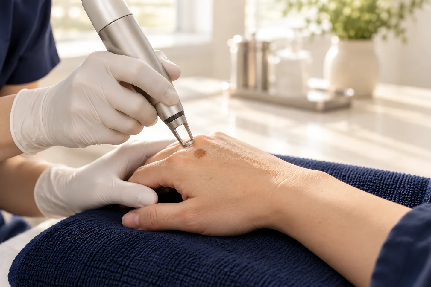
Pigment lasers can be useful for conditions such as freckles, solar lentigines, Hori’s Nevus, Nevus of Ota, and selected other pigment disorders. However, there is no single “pigment laser” that is appropriate for every brown spot. The correct treatment depends on the diagnosis, the depth and type of pigment, the patient’s skin phototype, previous treatments, and the risk of Post-Inflammatory Hyperpigmentation (PIH).
This distinction is particularly important for Thai and Asian skin because treatment-induced pigmentation may occur more easily in darker skin phototypes. Pigment laser treatment can also be inappropriate for certain conditions such as active melasma or undiagnosed pigmented lesions.
The safest approach is therefore:
diagnose the pigmentation first → determine where the pigment is located → select an appropriate wavelength and laser type → use
suitable treatment parameters → provide careful aftercare and photoprotection.
Lasers can successfully lighten many pigmented lesions, but response depends on pigment depth and lesion type, and recurrence is possible.What Is a Pigment Laser?
A pigment laser is a laser designed to selectively deliver energy to melanin or melanin-containing structures in the skin.
Melanin absorbs light over a broad range of wavelengths. Different wavelengths penetrate the skin to different depths and interact differently with epidermal melanin.
This is why a laser that works well for a superficial freckle may not be the most appropriate laser for a deeper gray-blue pigmentation such as Hori’s Nevus.
Common pigment-targeting laser systems include:
- Q-Switched Nd:YAG Laser
- Q-Switched Alexandrite Laser
- Q-Switched Ruby Laser
- Picosecond Laser
- Selected other pigment-targeting laser systems
IPL is frequently used for pigmentation but is technically not a laser. It uses filtered broad-spectrum light rather than a single laser wavelength.
How Does Pigment Laser Work?
Pigment laser treatment is based on the principle of Selective Photothermolysis.
The goal is to deliver light energy that is preferentially absorbed by melanin while limiting unnecessary damage to the surrounding skin.
When melanin absorbs the laser energy, the pigment-containing structures are disrupted into smaller particles or damaged sufficiently for the body to gradually clear them.
Short-pulse systems such as Q-Switched and Picosecond lasers can deliver high-energy pulses over extremely short periods.
This allows pigment to be targeted while reducing prolonged heating of surrounding tissue.
However, laser treatment still produces a controlled injury. If the inflammatory response is excessive, the skin may respond by creating more pigment, resulting in PIH.
What Does “Pigment Depth” Mean?
One of the most important factors in choosing a laser is where the pigment is located in the skin.
Pigmentation may be predominantly:
- Epidermal — closer to the skin surface
- Dermal — deeper within the skin
- Mixed — involving more than one level
Superficial pigment is generally more accessible to shorter wavelengths, while longer wavelengths can penetrate more deeply.
This is one reason why diagnosing the condition is more useful than simply choosing a laser based on its brand name.
What Can Pigment Laser Treat?
Depending on the laser and the diagnosis, pigment-targeting lasers may be considered for:
- Freckles
- Solar Lentigines
- Selected sunspots
- Hori’s Nevus
- Nevus of Ota
- Café-au-lait Macules in selected cases
- Certain benign melanocytic lesions
- Selected Post-Inflammatory Hyperpigmentation
- Selected resistant pigmentation disorders
Not all of these conditions respond in the same way.
Some superficial lesions may improve relatively quickly, while deeper dermal pigmentation often requires multiple treatments.
Other pigment disorders, particularly melasma and PIH, require considerably more caution.
Pigment Laser for Freckles
Freckles, medically known as Ephelides, are small superficial brown spots strongly influenced by genetics and ultraviolet exposure.
Because much of the pigment is relatively superficial, pigment-targeting lasers can be effective in appropriately selected patients.
Possible options include:
- 532 nm pigment lasers
- Q-Switched lasers
- Picosecond lasers
- Selected IPL systems
Clinical studies have shown that different short-pulse wavelengths can improve freckles, but treatment response and side effects vary according to wavelength, device, skin type, and treatment settings.
Importantly, laser does not remove the genetic tendency to develop freckles.
New freckles may appear after future ultraviolet exposure.
Pigment Laser for Solar Lentigines and Sunspots
Solar Lentigines are persistent brown spots caused largely by accumulated sun exposure.
They are particularly common on:
- Face
- Cheeks
- Hands
- Forearms
- Upper chest
- Other chronically sun-exposed areas
Several laser and light-based treatments can improve solar lentigines.
A recent systematic review of clinical trials found evidence supporting several procedural treatments, including Q-Switched lasers, Picosecond lasers, and IPL, although outcomes differed substantially across studies and treatment protocols.
Before treating an isolated sunspot, however, the lesion should be assessed to make sure it is truly benign.
Pigment Laser for Hori’s Nevus
Hori’s Nevus, also called Acquired Bilateral Nevus of Ota-Like Macules, is a dermal pigmentation disorder seen particularly in Asian patients.
It usually appears as symmetrical:
- Gray-brown spots
- Slate-gray spots
- Blue-gray spots
often affecting the cheekbones.
Unlike superficial freckles, the pigment in Hori’s Nevus is located deeper in the dermis.
Q-Switched and other short-pulse lasers have been used extensively for this condition. A systematic review found evidence for several laser approaches, including Alexandrite and Nd:YAG systems, but also noted differences in efficacy and risk of hyperpigmentation between treatment protocols.
Multiple treatment sessions are commonly required.
Pigment Laser for Nevus of Ota
Nevus of Ota is a form of dermal melanocytosis that usually produces blue-gray or brown pigmentation around the forehead, temple, cheek, or eye area.
Because the pigment is located within the dermis, longer-wavelength Q-Switched lasers such as 1064 nm Nd:YAG have traditionally been used.
Studies in darker Asian skin phototypes support the ability of Q-Switched Nd:YAG systems to improve Nevus of Ota, although multiple sessions may be required and pigmentary side effects can occur.
Pigmentation around the eye requires particularly careful assessment and appropriate eye protection during treatment.
Can Pigment Laser Treat Post-Inflammatory Hyperpigmentation?
Sometimes, but laser is usually not the first treatment for PIH.
Post-Inflammatory Hyperpigmentation develops after inflammation or injury, such as:
- Acne
- Eczema
- Burns
- Scratching
- Insect bites
- Chemical Peels
- Laser procedures
- Other skin injuries
This creates a particular problem: laser treatment itself can produce inflammation, and that inflammation can potentially create more PIH.
A systematic review focusing on skin of color found that laser treatment can improve PIH in some patients but also documented cases where pigmentation worsened after treatment.
Reviews therefore generally position laser as a second-line or adjunctive treatment for resistant PIH rather than a universal first treatment.Can Pigment Laser Treat Melasma?
Pigment lasers can sometimes be incorporated into treatment for carefully selected patients with melasma, but melasma should not be treated like a collection of freckles or sunspots.
Melasma is a chronic, relapsing pigment disorder involving more than simply excess visible melanin.
Aggressive pigment laser treatment can potentially lead to:
- Temporary improvement followed by recurrence
- PIH
- Rebound pigmentation
- Mottled hypopigmentation
- Uneven skin color
Therefore, photoprotection and appropriate medical treatment remain fundamental.
Laser treatment should generally be considered selectively rather than automatically because a patient has facial pigmentation.
This distinction is especially important in Asian skin.
What Is a Q-Switched Laser?
Q-Switched lasers generate very short nanosecond pulses with high peak energy.
These pulses can selectively disrupt pigment-containing structures.
Common Q-Switched pigment wavelengths include:
- 532 nm
- 694 nm Ruby
- 755 nm Alexandrite
- 1064 nm Nd:YAG
Each behaves differently within the skin.
Q-Switched systems have a long history of use for epidermal and dermal pigmentation and remain important tools for several pigment disorders.What Is a Picosecond Laser?
Picosecond Laser systems produce pulses measured in picoseconds, which are shorter than the nanosecond pulses produced by traditional Q-Switched systems.
Shorter pulses can produce stronger photoacoustic effects on pigment particles while limiting prolonged thermal exposure.
Depending on the device, wavelengths may include:
- 532 nm
- 730 nm
- 755 nm
- 785 nm
- 1064 nm
Picosecond lasers have demonstrated efficacy for several pigmentation conditions, including freckles and solar lentigines.
However,
“Picosecond” does not automatically mean safer, better, or more effective for every patient.
Wavelength, fluence, spot size, pulse characteristics, diagnosis, skin type, and clinical technique all matter.
Pico Laser vs Q-Switched Laser
Patients frequently ask whether a Pico Laser is better than a Q-Switched Laser.
There is no universal answer.
Both can be effective pigment-targeting technologies.
Picosecond Laser
Potential advantages may include efficient photoacoustic fragmentation and the availability of newer wavelength options.
Q-Switched Laser
Has extensive clinical experience and remains well established for many epidermal and dermal pigment disorders.
A newer machine does not necessarily compensate for an incorrect diagnosis or unsuitable treatment parameters.
For pigmentation, the question should therefore not simply be:
“Which machine is newer?”
A more useful question is:
“Which wavelength and treatment strategy are appropriate for this particular pigment disorder and skin type?”
What Does 532 nm Mean?
The number represents the laser wavelength.
532 nm is strongly absorbed by melanin and is frequently used for relatively superficial epidermal pigmentation.
Examples may include selected:
- Freckles
- Solar Lentigines
- Superficial pigment spots
Because 532 nm energy also interacts more strongly with epidermal melanin, it must be used carefully in darker skin phototypes.
The risk of unwanted pigment alteration depends on the device, pulse characteristics, treatment settings, and individual patient.
What Does 1064 nm Mean?
1064 nm Nd:YAG penetrates deeper into the skin and interacts less strongly with superficial epidermal melanin than shorter wavelengths.
This makes it useful for certain deeper pigment disorders and is one reason it is frequently considered in darker skin phototypes.
Examples may include:
- Nevus of Ota
- Hori’s Nevus
- Selected dermal pigment disorders
However, 1064 nm is not automatically safe simply because the wavelength is longer.
Incorrect parameters can still produce:
- PIH
- Hypopigmentation
- Burns
- Scarring
- Uneven pigmentation
What About 755 nm Alexandrite Laser?
The 755 nm Alexandrite wavelength can target melanin and has applications for both superficial and deeper pigmentation depending on the device and pulse characteristics.
Q-Switched and Picosecond Alexandrite systems have been studied for conditions including:
- Freckles
- Solar Lentigines
- Hori’s Nevus
- Other melanocytic pigmentation
Again, the suitability of the wavelength depends on the patient’s diagnosis and skin type.
What About Ruby Laser?
The 694 nm Ruby Laser has strong melanin absorption and has historically been used for several pigmented lesions.
Q-Switched Ruby Laser has been studied for:
- Freckles
- Solar Lentigines
- Hori’s Nevus
- Selected benign melanocytic lesions
Because of significant absorption by epidermal melanin, treatment requires caution in darker skin types.
Is IPL a Pigment Laser?
Technically, no.
Intense Pulsed Light (IPL) is a light-based device rather than a laser.
A laser generally produces a more specific wavelength, whereas IPL produces a range of wavelengths that are filtered according to the treatment target.
IPL can nevertheless be useful for certain superficial pigment problems, particularly when multiple sunspots are spread over a larger area.
IPL may also simultaneously influence vascular and photodamage-related changes depending on the system.
It should not, however, be used indiscriminately for all forms of pigmentation.
Which Pigment Laser Is Best?
There is no single pigment laser that is best for everyone.
Choosing treatment requires considering:
- Diagnosis
- Pigment color
- Pigment depth
- Skin phototype
- Location
- Size and number of lesions
- Previous treatments
- Previous PIH
- Recent sun exposure
- Desired downtime
- Treatment goals
A superficial brown freckle and a deep gray Hori’s Nevus may look like “pigmentation” to a patient but require very different treatment strategies.
Why Diagnosis Is More Important Than the Laser Brand
Many patients search for a particular device before knowing what type of pigmentation they actually have.
This can lead to inappropriate treatment.
For example:
- Freckles may respond well to targeted pigment treatment
- Solar Lentigines may respond well but should first be confirmed as benign
- Hori’s Nevus usually requires treatment suitable for deeper dermal pigment
- Melasma can worsen after inappropriate laser treatment
- PIH may worsen if additional inflammation is created
- A suspicious mole should not simply be destroyed with a cosmetic laser
The correct diagnosis should therefore come before selecting a device.
Should Every Brown Spot Be Treated With Laser?
No.
This is one of the most important safety principles of pigmentation treatment.
If a pigmented lesion is:
- New
- Changing
- Asymmetrical
- Irregularly bordered
- Multicolored
- Growing
- Bleeding
- Ulcerated
- Persistently crusting
- Significantly different from surrounding spots
it should be medically assessed rather than automatically treated with a pigment laser.
Medical guidelines specifically advise that pigmented lesions with atypical features should be biopsied to exclude malignancy before laser treatment.
Destroying an undiagnosed lesion can remove useful diagnostic information and delay appropriate treatment.
How Does a Dermatologist Evaluate Pigmentation Before Laser?
Assessment may include:
- Clinical examination
- Distribution of pigmentation
- Color of the lesion
- Depth suggested by appearance
- Whether lesions are symmetrical
- Skin phototype
- History of sun exposure
- Previous pigmentation
- Previous laser treatment
- History of PIH
- Medications
- Recent tanning
- Active acne or dermatitis
- History of abnormal scarring
Dermoscopy may be useful for selected lesions.
A Skin Biopsy may be considered when the diagnosis is uncertain or a lesion has suspicious features.
Why Is Pigment Laser Different in Asian Skin?
Asian skin generally contains more epidermal melanin than very fair skin.
That melanin can compete with the pigmentation being targeted for absorption of laser energy.
This increases the importance of:
- Correct wavelength selection
- Conservative treatment parameters when appropriate
- Avoiding unnecessary overlap
- Avoiding treatment of recently tanned skin
- Appropriate cooling when relevant
- Photoprotection
- Monitoring for PIH
Reviews of laser treatment in Asian skin emphasize a greater susceptibility to unwanted pigment changes following laser procedures.
Post-Inflammatory Hyperpigmentation After Pigment Laser
PIH is one of the most important complications of pigment laser treatment in Asian and darker skin.
Following laser treatment, inflammation can stimulate Melanocyte activity and increase melanin production.
As a result, a treated spot may initially improve but later become darker.
Risk can be influenced by:
- Skin phototype
- Treatment intensity
- Depth of injury
- Recent sun exposure
- Existing inflammation
- Previous PIH
- Picking or scratching
- Inadequate photoprotection
- Individual biological response
PIH is especially common following inflammatory procedures in darker skin phototypes.Can PIH Be Prevented?
There is no method that completely eliminates the risk.
However, appropriate treatment selection and careful photoprotection are important.
A 2025 systematic review examining prevention of PIH in skin of color found that sunscreen was the intervention most consistently associated with preventing post-procedure PIH, although available evidence remains limited.
Practical prevention includes:
- Avoiding unnecessary sun exposure
- Using appropriate sunscreen
- Avoiding treatment on recently tanned skin
- Using appropriate laser parameters
- Controlling active inflammation
- Avoiding picking or scratching after treatment
- Following post-procedure skincare instructions
Can Pigment Laser Cause Hypopigmentation?
Yes.
Hypopigmentation means the treated area becomes lighter than the surrounding skin.
This may occur if pigment-producing cells are excessively affected by treatment.
It may be:
- Temporary
- Prolonged
- Rarely permanent
It is specifically noted that permanent hypopigmentation or depigmentation can occur with pigment laser treatment, particularly in skin of color.
Can Pigment Laser Cause Scarring?
Yes, although scarring should be uncommon with appropriate treatment.
Risk may increase with:
- Excessive energy
- Excessive treatment overlap
- Burns
- Significant blistering
- Infection
- Picking or removing crusts
- Abnormal wound healing
Patients with a history of Keloid or other abnormal scars should inform their dermatologist before treatment.
Other Possible Side Effects
Depending on the device and intensity, temporary effects may include:
- Redness
- Swelling
- Stinging
- Burning sensation
- Tenderness
- Darkening of treated pigment
- Fine crusting
- Mild bruising
- Temporary sensitivity
Less common but more significant complications include:
- PIH
- Hypopigmentation
- Blistering
- Infection
- Prolonged redness
- Persistent pigment alteration
- Scarring
Does Pigmentation Get Darker Immediately After Laser?
It can.
Certain superficial pigmented lesions may temporarily become darker after treatment.
This can represent treated pigment and superficial tissue changes rather than treatment failure.
Depending on the procedure, the darkened area may gradually flake or fade.
Patients should generally avoid scraping, peeling, or picking treated spots.
What Happens During Pigment Laser Treatment?
The exact process varies by device and condition, but commonly includes:
- Diagnosis and skin assessment
The dermatologist confirms whether the pigmentation is appropriate for laser treatment.
- Photography or documentation
Baseline documentation can be useful for comparing treatment response over time.
- Skin preparation
The treatment area is cleaned and eye protection is provided.
- Laser treatment
Energy is delivered to selected pigmented areas using parameters appropriate for the lesion and skin type.
- Immediate skin response
Depending on the laser, the treated area may show:
- Temporary whitening
- Mild redness
- Darkening
- Swelling
- Pinpoint changes
- Aftercare
The patient receives instructions regarding cleansing, skincare, sun protection, and expected recovery.
Does Pigment Laser Hurt?
Discomfort depends on:
- Laser type
- Wavelength
- Fluence
- Spot size
- Treatment area
- Number of lesions
- Individual sensitivity
Patients may describe:
- Stinging
- Heat
- Pricking
- A sensation similar to a small elastic band snapping against the skin
Depending on the procedure, cooling or local anesthesia may be considered.
How Long Is Recovery?
Recovery varies considerably because “pigment laser” includes many different technologies.
Some treatments may produce only mild redness for a short period.
Others may cause:
- Darkening of spots
- Fine crusting
- Mild swelling
- Temporary tenderness
- Several days of visible healing
The expected recovery should therefore be discussed for the specific laser and lesion being treated rather than generalized to every pigment procedure.
Aftercare After Pigment Laser
General aftercare may include:
- Clean the skin gently
- Avoid scrubbing
- Avoid picking crusts
- Use moisturizer as recommended
- Pause irritating active skincare when appropriate
- Avoid excessive heat if the skin remains inflamed
- Protect the treated area from sunlight
- Use broad-spectrum sunscreen
- Follow device-specific instructions
For patients who develop blisters, increasing pain, pus, spreading redness, or delayed healing, medical review is appropriate.
When Can Skincare Be Restarted?
This depends on the intensity of treatment and how quickly the skin barrier recovers.
Products that may temporarily need to be paused include:
- Retinoids
- Strong exfoliating acids
- Scrubs
- Irritating acne treatments
- Other potentially sensitizing products
They can usually be restarted when the skin has recovered according to the treating clinician’s instructions.
How Many Pigment Laser Sessions Are Needed?
There is no universal answer.
The number of treatments may depend on:
- Diagnosis
- Pigment depth
- Pigment density
- Size
- Number of lesions
- Laser type
- Wavelength
- Skin phototype
- Treatment parameters
- Individual response
- Risk of PIH
A superficial sunspot and Hori’s Nevus should not be expected to require the same treatment schedule.
Patients should be cautious of guarantees that all pigmentation can be removed permanently in a predetermined number of sessions.
How Long Should You Wait Between Sessions?
Treatment intervals depend on the diagnosis and laser technique.
A dermatologist may need to allow time for:
- Pigment clearance
- Resolution of inflammation
- PIH assessment
- Skin barrier recovery
- Evaluation of treatment response
Repeating treatment simply because pigment remains visible immediately after a session may increase unnecessary inflammation.
Can Pigmentation Return After Laser?
Yes.
Recurrence depends on the original condition.
Freckles
Can reappear or new freckles can develop after ultraviolet exposure.
Solar Lentigines
New lesions may appear as cumulative sun damage continues.
Melasma
Recurrence is common because the underlying biological tendency persists.
Hori’s Nevus and Nevus of Ota
Response and recurrence patterns differ from superficial sun-related pigmentation and often involve longer treatment courses.
Successfully treated pigmented lesions can recur.
Does Pigment Laser Permanently Remove Pigmentation?
Sometimes a particular benign lesion can achieve long-lasting clearance, but permanent removal cannot be guaranteed for pigmentation as a category.
The result depends on the condition.
Laser can remove existing pigment without changing:
- Genetic predisposition
- Future sun exposure
- Hormonal triggers
- Skin inflammation
- Aging
- The tendency to produce melanin
Realistic expectations are therefore essential.
Who May Be a Good Candidate?
Pigment laser may be considered for patients who:
- Have a clearly diagnosed benign pigment disorder
- Want cosmetic improvement
- Understand that multiple sessions may be needed
- Can follow sun protection recommendations
- Do not have active inflammation in the treatment area
- Understand the possibility of recurrence and pigmentary side effects
The final decision depends on the specific diagnosis.
When Should Pigment Laser Be Delayed or Avoided?
Treatment may need to be postponed, modified, or avoided when there is:
- Uncertain diagnosis
- Suspicious pigmented lesion
- Recent significant tanning
- Sunburn
- Active skin infection
- Open wounds
- Active dermatitis
- Significant skin irritation
- Poor wound healing
- Previous serious reaction to laser
- Other relevant medical contraindications
Pregnancy and medication history may also affect treatment planning depending on the procedure and associated medications.
Is a Test Spot Necessary?
A test spot can sometimes be useful, particularly when:
- The skin phototype is darker
- The pigment diagnosis is unusual
- The patient has a history of PIH
- The planned treatment is relatively aggressive
- The response of the lesion is uncertain
However, a test spot cannot guarantee that PIH or another complication will not occur elsewhere.
Why Recent Tanning Matters
Recent ultraviolet exposure increases melanin in the epidermis.
This can increase competition between normal skin pigment and the intended target.
Potential consequences include:
- Greater epidermal heating
- Burns
- PIH
- Hypopigmentation
- Less predictable response
Patients should tell their dermatologist about recent beach holidays, tanning, or significant outdoor exposure before treatment.
Pigment Laser and Sun Protection in Bangkok
Sun protection is particularly important when undergoing pigmentation treatment in Bangkok because ultraviolet exposure occurs throughout the year.
Patients should consider:
- Daily sunscreen
- Adequate application
- Reapplication during prolonged outdoor exposure
- Hats
- Shade
- Avoiding intentional tanning
- Additional protection during outdoor activities
Sun protection does not guarantee that pigmentation will never return, but it helps reduce ultraviolet stimulation and treatment-related pigment problems.
Pigment Laser for Tourists and International Patients in Bangkok
Patients visiting Bangkok specifically for pigmentation treatment should consider their travel itinerary before undergoing a procedure.
Important questions include:
- How long will you remain in Bangkok?
- Are you travelling to a beach afterward?
- Will you be swimming or diving?
- Will you spend long periods outdoors?
- Do you have an important event soon?
- Will visible crusting or darkening be acceptable?
- Could follow-up be needed?
- Will multiple sessions be required?
Thailand is a popular destination for beaches and outdoor activities, which can conflict with post-laser sun protection.
For some patients, it may be more practical to schedule treatment toward a period when strict aftercare is possible rather than immediately before an island or beach holiday.
When Should You See a Dermatologist Before Pigment Laser?
Dermatology assessment is particularly important if:
- You do not know what type of pigmentation you have
- There are several different kinds of brown or gray spots
- One lesion looks different from the others
- A lesion has changed
- Pigmentation has worsened after previous laser treatment
- You have melasma
- You have a history of significant PIH
- You have darker skin and are concerned about laser complications
- You have previously developed hypopigmentation
- You are considering repeated laser treatments
- A spot bleeds, ulcerates, crusts, grows, or changes color
Warning Signs That Should Be Checked Before Cosmetic Laser Treatment
Do not simply laser a pigmented lesion if it has concerning features such as:
- New and rapidly changing
- Asymmetrical appearance
- Irregular border
- Multiple uneven colors
- Progressive growth
- Bleeding
- Ulceration
- Recurrent crusting
- Persistent itching or pain
- A distinctly different appearance from surrounding lesions
Such lesions may require Dermoscopy, clinical follow-up, or Skin Biopsy rather than cosmetic destruction.
What to Expect During a Pigmentation Consultation in Bangkok
A useful pigmentation consultation should focus first on diagnosis rather than selecting a machine.
The dermatologist may assess:
- Whether the pigmentation is epidermal, dermal, or mixed
- Whether the diagnosis is freckles, solar lentigines, melasma, PIH, Hori’s Nevus, Nevus of Ota, or another disorder
- Skin phototype
- Pigment color and distribution
- Sun exposure
- Recent tanning
- Previous lasers
- Previous PIH
- Previous hypopigmentation
- Active acne or dermatitis
- Current skincare
- Medications
- Suspicious lesions requiring further assessment
Only after this evaluation should the wavelength, device, energy, treatment strategy, and expected number of treatments be considered.
Frequently asked questions
What is Pigment Laser?
Pigment Laser is a general term for lasers designed to target melanin-containing pigmentation in the skin.
What conditions can Pigment Laser treat?
Depending on the device, it may be used for freckles, solar lentigines, Hori’s Nevus, Nevus of Ota, and selected other pigment disorders.
Is Pigment Laser suitable for every brown spot?
No. Different brown spots have different causes and depths, and some lesions should not be treated with laser.
What is the best laser for pigmentation?
There is no single best laser. The appropriate wavelength and device depend on the diagnosis, pigment depth, skin phototype, and risk of complications.
Is Pico Laser the same as Pigment Laser?
Pico Laser is one category of laser technology that can be used for pigmentation. Pigment Laser is a broader term.
Is Q-Switched Laser a Pigment Laser?
Yes. Q-Switched systems are widely used for both superficial and dermal pigmented lesions.
Is IPL a laser?
No. IPL uses filtered broad-spectrum light rather than a single laser wavelength.
What does 532 nm laser treat?
It is commonly used for selected superficial pigmentation such as freckles and solar lentigines.
What does 1064 nm laser treat?
The wavelength penetrates more deeply and may be used for selected dermal pigmentation such as Hori’s Nevus and Nevus of Ota.
Is 1064 nm safer for darker skin?
It generally interacts less strongly with superficial epidermal melanin than shorter wavelengths, but it can still cause burns, PIH, hypopigmentation, and other complications if used inappropriately.
Can Pigment Laser remove freckles?
Yes, freckles can often be lightened significantly, but new freckles may appear after future sun exposure.
Can Pigment Laser remove sunspots?
Selected solar lentigines respond well to pigment-targeting lasers and light-based treatments, but correct diagnosis is important before treatment.
Can Pigment Laser treat Hori’s Nevus?
Yes. Q-Switched and other short-pulse laser treatments have evidence supporting their use for Hori’s Nevus, although several sessions may be required.
Can Pigment Laser treat Nevus of Ota?
Yes. Short-pulse lasers, including Q-Switched Nd:YAG, are established treatments for Nevus of Ota.
Can Pigment Laser treat PIH?
Sometimes, but laser is generally considered an adjunct or second-line option because treatment itself can worsen PIH.
Can Pigment Laser treat melasma?
It may have a role in selected patients, but melasma requires particular caution because laser can trigger rebound pigmentation, PIH, or recurrence.
Can laser make pigmentation darker?
Yes. PIH can develop after laser-induced inflammation, particularly in darker skin phototypes.
Can laser make the skin white?
Excessive damage to pigment-producing cells can cause Hypopigmentation or, rarely, prolonged depigmentation.
Is Pigment Laser safe for Asian skin?
It can be used safely in appropriately selected patients, but Asian skin has an important risk of treatment-induced pigment changes, making diagnosis and parameter selection particularly important.
Does Pigment Laser hurt?
Treatment may feel like stinging, heat, or small elastic-band snaps. Discomfort depends on the device, treatment parameters, and location.
Do I need anesthesia?
Some procedures do not require it. Depending on the treatment, cooling or local anesthesia may be considered.
What does the skin look like immediately after treatment?
Depending on the laser, there may be redness, temporary whitening, mild swelling, or darkening of the pigmented spots.
Why do spots get darker after laser?
Temporary darkening can occur as treated pigment and superficial tissue respond to laser energy before gradually clearing.
Should I peel off dark spots after laser?
No. Picking or peeling can increase the risk of PIH, infection, hypopigmentation, and scarring.
How long does recovery take?
It varies by device and treatment intensity. Some procedures cause minimal visible recovery, while others produce several days of darkening or crusting.
How many sessions will I need?
There is no fixed number. Superficial freckles may behave very differently from deeper dermal pigmentation such as Hori’s Nevus.
Can pigmentation come back?
Yes. Recurrence or development of new pigmentation is possible, particularly with continued sun exposure or conditions such as melasma and freckles.
Is Pigment Laser permanent?
Permanent clearance cannot be guaranteed for pigmentation in general. Long-term results depend strongly on the underlying diagnosis.
Can I have Pigment Laser after going to the beach?
Recent tanning can increase treatment risks. A dermatologist should assess the skin before treatment.
Can I go to the beach after Pigment Laser?
Immediate strong ultraviolet exposure is generally undesirable during healing and may increase pigment complications.
Should I use sunscreen after Pigment Laser?
Yes. Photoprotection is an important part of reducing treatment-related PIH and recurrent pigmentation.
Can every mole be removed with Pigment Laser?
No. Moles and unusual pigmented lesions should be diagnosed first. Suspicious lesions may need Skin Biopsy rather than laser destruction.
Can Pigment Laser hide skin cancer?
Treating an undiagnosed suspicious pigmented lesion can alter its appearance and delay diagnosis. Such lesions should be assessed before cosmetic treatment.
Should I see a dermatologist before Pigment Laser?
It is particularly advisable when the diagnosis is uncertain, pigmentation is mixed, a lesion is changing, you have melasma or significant PIH, or previous laser treatment caused pigment complications.
Related conditions
-
Freckles Freckles Freckles, medically called ephelides, are small, flat, light-brown to darker-brown spots that commonly appear on sun-exposed skin, especially the face, nose, cheeks, shoulders, arms, and upper chest.
-
Melasma Melasma Melasma is a common acquired pigmentation disorder that causes symmetrical brown, gray-brown, or sometimes darker patches, most often on the cheeks, forehead, nose, upper lip, and jawline.

-
Sun Spots Sun Spots A sun spot is a common term for a flat brown mark that develops on sun-exposed skin. In dermatology, many of these spots are solar lentigines—benign areas of increased pigmentation associated with cumulative sun exposure.

-
Post-inflammatory Hyperpigmentation Post-inflammatory Hyperpigmentation Post-inflammatory hyperpigmentation (PIH) is the flat brown, dark brown, or grayish discoloration that remains after inflammation or injury to the skin. It commonly follows acne, eczema, insect bites, burns, scratching, irritation, and some dermatologic procedures.