Mole Removal
Many growths that patients call “moles” are benign and can be removed because they catch on clothing, become repeatedly irritated, or are cosmetically unwanted. Others may actually be Seborrheic Keratosis, Skin Tag, Wart, Actinic Keratosis, or another skin condition. Occasionally, a lesion that looks like an ordinary mole or growth can represent Melanoma, Basal Cell Carcinoma, SCC, or another skin cancer.
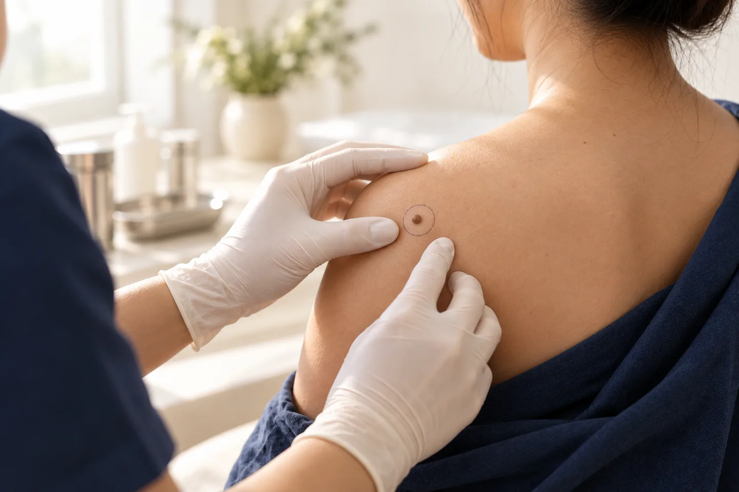
Mole and skin lesion removal should begin with one question: what is the lesion?
Many growths that patients call “moles” are benign and can be removed because they catch on clothing, become repeatedly irritated, or are cosmetically unwanted. Others may actually be Seborrheic Keratosis, Skin Tag, Wart, Actinic Keratosis, or another skin condition. Occasionally, a lesion that looks like an ordinary mole or growth can represent Melanoma, Basal Cell Carcinoma, SCC, or another skin cancer.
Most benign moles do not need treatment. When removal is appropriate, common approaches include Shave Removal and Surgical Excision. Suspicious lesions require a different approach: obtaining an adequate specimen for Histopathology takes priority over simply choosing the quickest or least invasive cosmetic technique.
For Thai patients, expats, tourists, and international patients looking for Mole Removal in Bangkok, the safest treatment pathway is:
Skin lesion assessment → diagnosis or Dermoscopy when appropriate → decide whether Histopathology is needed → select the appropriate removal method → wound care → review pathology when required.
What Is a Mole?
A mole, medically called a melanocytic nevus, is a benign growth of melanocytes, the pigment-producing cells in the skin.
Moles can differ considerably in appearance. They may be flat or raised, smooth or slightly rough, and can be skin-colored, pink, tan, brown, dark brown, or occasionally very dark. Some contain hair.
A typical benign mole tends to have a relatively regular shape, consistent color, and remains fairly stable over time. Most moles require no treatment unless they are bothersome, cosmetically unwanted, or suspicious for skin cancer.
Not Every Skin Growth Is a Mole
One of the most useful things a dermatologist can do before removal is determine what type of lesion is actually present.
Conditions commonly mistaken for moles include:
- Seborrheic Keratosis
- Skin Tag
- Dermatosis Papulosa Nigra
- Wart
- Solar Lentigo
- Actinic Keratosis
- Dermatofibroma
- Cyst
- benign melanocytic nevus
- Basal Cell Carcinoma
- SCC
- Melanoma
This distinction matters because these lesions are treated differently.
For example, a typical Seborrheic Keratosis may be treated with Cryotherapy, Curettage, Electrocautery, Shave Removal, or selected other methods. A suspicious pigmented lesion may instead require Biopsy or complete surgical removal so that tissue remains available for Histopathology.
Why Should a Mole or Skin Lesion Be Checked Before Removal?
The main reason is that cosmetic destruction and diagnostic removal are not the same thing.
If a lesion is confidently benign, treatment can focus on symptoms, cosmetic outcome, wound healing, recurrence, and scar risk.
If the diagnosis is uncertain or Melanoma or another skin cancer is possible, the priority changes. The dermatologist needs to choose a procedure that provides enough tissue for an accurate microscopic diagnosis.
This is particularly important before destructive treatments that may leave little or no intact specimen for Histopathology.
A useful principle is:
Diagnosis first. Removal method second.
How Does a Dermatologist Assess a Mole?
Assessment usually combines the patient’s history and clinical examination.
The dermatologist may consider:
- how long the lesion has been present;
- whether it is new or changing;
- whether it bleeds or repeatedly crusts;
- change in size, shape, or color;
- whether it is painful or itchy;
- symmetry and border;
- color pattern;
- whether it looks different from the patient’s other moles;
- previous treatment or removal;
- history of Melanoma or other skin cancer.
For pigmented lesions, Dermoscopy may help the dermatologist see structures and pigmentation patterns below the surface that cannot be appreciated as clearly with the naked eye.
Dermoscopy can improve clinical assessment, but Histopathology remains necessary when tissue diagnosis is required.
What Are the Warning Signs of Melanoma?
The ABCDE rule is a useful guide for identifying suspicious pigmented lesions:
A — Asymmetry
One half of the lesion looks different from the other.
B — Border
The border is irregular, scalloped, notched, or poorly defined.
C — Color
There are multiple or uneven colors rather than a consistent pigment pattern.
D — Diameter
Melanomas are often larger than about 6 mm when diagnosed, but they can be smaller, so diameter alone should never be used to rule Melanoma in or out.
E — Evolving
The lesion is changing in size, shape, color, or overall appearance.
A spot that looks distinctly different from the patient’s other moles—the so-called ugly duckling—also deserves attention. New or changing lesions, or spots that itch or bleed, should be assessed by a dermatologist.
Other Skin Cancer Warning Signs
Not every skin cancer looks like a dark mole.
Skin cancer may also present as:
- a growing bump;
- a lesion that repeatedly bleeds;
- a rough or scaly patch;
- a firm growth;
- a shiny bump;
- a sore that does not heal;
- a sore that appears to heal but returns;
- a persistent “pimple” or “insect bite” that does not go away.
A lesion with these features should be evaluated rather than repeatedly destroyed or removed purely for cosmetic reasons.
Does Every Mole Need to Be Removed?
No.
Most benign moles can safely remain untreated.
Mole Removal is generally considered for three broad reasons.
Cosmetic Removal
A benign mole may be removed because the patient does not like its appearance.
Before cosmetic removal, the dermatologist should discuss whether the expected scar, depression, pigmentation change, or recurrence might be more noticeable than the original mole.
Recurrent Irritation
A raised mole may repeatedly catch on:
- clothing;
- necklaces;
- bra straps;
- shaving blades;
- combs;
- other areas of friction.
Irritation and cosmetic concerns are recognized reasons for removing a benign mole.
Diagnostic Removal
If Melanoma or another skin cancer needs to be excluded, the purpose is no longer simply cosmetic.
The removal technique should provide adequate tissue for Histopathology.
Main Mole Removal Methods
For melanocytic moles, the two most important surgical approaches are:
Shave Removal and Surgical Excision.
Which one is more appropriate depends on:
- whether the mole is raised or flat;
- suspected diagnosis;
- lesion depth;
- location;
- cosmetic goals;
- importance of complete removal;
- recurrence risk;
- need for Histopathology.
Shave Mole Removal
Shave Removal, Shave Excision, or Tangential Excision involves using a surgical blade to remove a protruding lesion horizontally.
It is particularly suitable for selected lesions that project above the surrounding skin, including papillomatous melanocytic nevi and several other superficial growths.
How Is Shave Removal Performed?
The skin is usually numbed with local anesthesia.
The dermatologist then uses a blade to remove the elevated portion of the lesion. Bleeding is controlled, and many superficial wounds are allowed to heal naturally by Secondary Intention, meaning new tissue gradually fills the wound from the base.
A standard superficial Shave Biopsy generally does not require stitches.
Advantages of Shave Removal
For appropriately selected benign raised lesions, advantages may include:
- relatively straightforward procedure;
- local anesthesia;
- usually no sutures;
- suitable for raised lesions;
- tissue can still be sent for Histopathology.
Limitations of Shave Removal
The main limitation is that the deeper portion of the lesion may remain.
This means a melanocytic mole can grow back after Shave Removal.
The recurrent area may develop pigmentation within the scar and occasionally look irregular, which can complicate later assessment.
Therefore, Shave Removal is not automatically the best technique simply because it avoids stitches.
Deep Shave or Saucerization
A Deep Shave or Saucerization Biopsy removes a deeper, bowl-shaped portion of tissue.
It can provide more depth than a superficial shave and is sometimes used when deeper diagnostic information is necessary.
Because it removes more tissue, the resulting defect and scar can also be more noticeable, and some deeper wounds may require sutures.
Surgical Excision for Mole Removal
Surgical Excision removes the lesion through the full thickness of the skin.
The wound is generally closed with sutures.
Excision can be particularly useful when:
- a mole is flat rather than protruding;
- complete removal is desirable;
- the lesion extends deeper into the skin;
- the lesion has previously recurred after partial removal;
- Histopathology of the complete lesion is important;
- Melanoma or another significant diagnosis needs to be excluded.
Excision specimens can also allow the pathologist to assess the lesion and, when appropriate, whether the lesion reaches the examined surgical margins.
Shave Removal vs Surgical Excision
Neither method is universally better.
Shave Removal may be preferable when:
The lesion is clearly benign, protruding, and superficial, and the patient understands that recurrence or residual pigmentation can occur.
Surgical Excision may be preferable when:
Complete removal is important, the lesion is flat or deeper, recurrent disease would be problematic, or full-thickness Histopathology is needed.
The cosmetic trade-off differs.
Shave Removal may leave a round, slightly depressed or differently colored scar.
Excision usually creates a longer linear scar because the skin must be shaped and sutured to close the defect.
A longer scar is not necessarily a worse scar. For some lesions, a predictable linear scar can be preferable to recurrent pigmentation or a depressed Shave scar.
Can a Suspicious Mole Be Shaved Off?
Sometimes a sufficiently deep shave can provide adequate diagnostic tissue, but a superficial cosmetic shave should not be chosen when Melanoma is a meaningful concern if it risks cutting through the deepest portion of the tumor.
If Melanoma is present, tumor depth can affect subsequent staging and treatment planning.
When a shave technique is used for a lesion suspicious for Melanoma, it must extend deeply enough beneath the suspicious area. A Deep Shave or Saucerization is preferred to a superficial shave when Melanoma risk is higher.
The correct biopsy depends on the individual lesion.
Excisional Biopsy for a Suspicious Mole
When clinically feasible, an Excisional Biopsy may be used to remove the entire suspicious lesion for diagnosis.
This gives the pathologist a more complete view of the architecture of the lesion.
If Melanoma is subsequently diagnosed, additional treatment—usually Wide Local Excision—may still be required according to the Histopathology.
The initial diagnostic removal and definitive Melanoma surgery should therefore not be confused.
Should Every Removed Mole Be Sent for Histopathology?
Histopathology is particularly important when a melanocytic lesion or diagnostically uncertain lesion is surgically removed.
Removed moles are commonly examined microscopically to check for cancer cells.
Histopathology can identify conditions such as:
- benign melanocytic nevus;
- atypical or dysplastic nevus;
- Melanoma;
- Basal Cell Carcinoma;
- SCC;
- Seborrheic Keratosis;
- other skin tumors.
The decision about Histopathology should ideally be made before treatment, especially when considering destructive techniques that may leave no useful specimen.
What Does a Histopathology Report Tell Us?
A skin specimen sent to a pathology laboratory is processed and examined under a microscope.
Histopathology may be used to:
- confirm the diagnosis;
- identify tumor type;
- evaluate microscopic characteristics;
- assess whether an excised lesion reaches the examined margin, when the specimen permits this.
Additional stains or specialist review may occasionally be needed for more complex lesions.
What If the Removed Mole Is Melanoma?
If Histopathology shows Melanoma, management changes from cosmetic Mole Removal to Melanoma treatment.
The report may contain information needed for further planning, including tumor depth.
Further surgery, usually Wide Local Excision, is generally required after Melanoma diagnosis. Selected patients may also require additional staging assessment.
A superficial biopsy that cuts through the bottom of Melanoma can make accurate depth measurement more difficult, which is why proper initial biopsy planning matters.
Can Basal Cell Carcinoma Look Like a Mole?
Yes.
Some Basal Cell Carcinomas contain pigment and may resemble a brown or black growth.
Other presentations include:
- shiny papules;
- slowly growing bumps;
- persistent sores;
- lesions that bleed or crust repeatedly.
Because Basal Cell Carcinoma and other skin cancers can have variable appearances, a growth that is new, persistent, changing, or repeatedly bleeding deserves clinical assessment.
Can SCC Be Mistaken for a Benign Skin Lesion?
Yes.
SCC may appear as a:
- rough or scaly patch;
- crusted lesion;
- firm bump;
- enlarging growth;
- non-healing ulcer.
A growth repeatedly treated as a wart,
“dry skin,
” or benign bump that continues to enlarge, ulcerate, or bleed should be reassessed.
If SCC is suspected, preserving tissue for Histopathology becomes more important than cosmetic destruction.
Seborrheic Keratosis Removal
Seborrheic Keratosis is a common benign skin growth that may be brown, tan, dark, rough, waxy, or appear as if it is “stuck onto” the surface.
If the diagnosis is clear, treatment may be considered because the lesion:
- catches on clothing;
- becomes itchy;
- becomes irritated;
- is cosmetically unwanted.
Treatment options can include:
- Cryotherapy;
- Curettage;
- Electrocautery;
- Shave Removal;
- selected ablative treatments.
Pigment loss is an important consideration after destructive treatment, particularly in patients with darker skin.
Skin Tag Removal
Skin Tags or Acrochordons are soft benign growths commonly found in areas where skin rubs against skin or clothing.
Common locations include:
- neck;
- armpits;
- groin;
- eyelids;
- skin folds.
Possible treatments include Cryotherapy, surgical excision or snipping, Electrosurgery, and ligation in selected circumstances.
However, Seborrheic Keratosis, warts, and Molluscum Contagiosum can resemble Skin Tags, so unusual-looking lesions should be assessed before removal.
Dermatosis Papulosa Nigra Removal
Dermatosis Papulosa Nigra (DPN) consists of multiple small dark papules, often on the face and neck.
It is benign and usually does not require treatment.
When removal is desired, options may include Curettage, Electrodessication, Cryotherapy, or selected laser procedures.
An important consideration is that destructive treatment can cause:
- Hyperpigmentation;
- Hypopigmentation;
- scarring;
- Keloid formation.
This is especially relevant when treating numerous facial lesions in more deeply pigmented skin.
Can Actinic Keratosis Be Removed?
Actinic Keratosis is a sun-damaged keratinocytic lesion and should not simply be grouped together with cosmetic moles.
Treatment depends on the number, thickness, location, and clinical features.
Individual lesions may sometimes be treated with Cryotherapy. Thick or hyperkeratotic lesions may require Shave Removal, Curettage, or Electrocautery.
A thick, tender, rapidly growing, ulcerated, or otherwise concerning lesion may need Biopsy to exclude SCC.
Can Mole Removal Be Done With Laser?
Laser can be used for selected benign pigmented or superficial lesions, but its role should depend on the diagnosis.
The main concern with destructive treatment of an uncertain pigmented lesion is loss of diagnostic tissue.
A lesion that could represent Melanoma should be evaluated and, when indicated, biopsied appropriately rather than simply treated until the pigment disappears.
Can Cryotherapy Be Used for Mole Removal?
Cryotherapy has a valuable role for several benign and precancerous lesions, but it is generally not the preferred technique for an undiagnosed melanocytic mole.
Cryotherapy destroys tissue rather than providing an intact specimen suitable for full Histopathology.
This makes diagnosis-first planning especially important for pigmented lesions.
Can Electrocautery or Curettage Remove Skin Lesions?
Yes, in appropriately selected superficial lesions.
These methods are commonly considered for certain:
- Seborrheic Keratoses;
- Skin Tags;
- Warts;
- superficial keratinocytic lesions.
They are not interchangeable with diagnostic Biopsy.
A lesion that is clinically uncertain should be diagnosed before it is destroyed.
Can a Mole Grow Back After Removal?
Yes.
Recurrence is particularly possible after partial removal.
With Shave Removal, nevus cells may remain deeper in the skin. The treated area can later develop pigment or become raised again.
Patients are advised to have a mole reassessed if it grows back after removal.
A recurrent mole is not automatically Melanoma, but it should not simply be repeatedly removed without considering the previous diagnosis and Histopathology.
What Is a Recurrent Nevus?
A Recurrent Nevus is a melanocytic lesion that reappears after partial removal.
It may present as pigmentation developing within or around the scar.
Because pigmentation can be irregular, the recurrent lesion may look more concerning than the original mole.
When possible, keeping the original Histopathology report is valuable because it helps future dermatologists understand what was previously removed.
Does Mole Removal Hurt?
Most minor Mole Removal procedures are performed using local anesthesia.
The injection may sting or burn briefly.
Once the area is numb, the patient may still feel pressure, pulling, or movement, but should not feel sharp pain from the procedure itself.
Tenderness after the anesthetic wears off varies according to the size, site, depth, and technique used.
Does Mole Removal Leave a Scar?
Yes. Any procedure that removes skin can leave a permanent mark.
There is no technique that can guarantee completely scar-free Mole Removal.
Depending on the procedure and individual healing, the treated area may become:
- a small flat mark;
- a round pale scar;
- a darker mark;
- a slightly depressed scar;
- a linear surgical scar;
- a Hypertrophic Scar;
- a Keloid.
Even small skin biopsies can leave permanent skin change.
For cosmetic lesions, the expected scar should be discussed before deciding whether removal is worthwhile.
Shave Scar vs Excision Scar
The two procedures tend to produce different types of scars.
A Shave Removal commonly creates a round wound that heals naturally. It may eventually become flat, slightly depressed, lighter, or darker than the surrounding skin.
A Surgical Excision usually produces a linear scar because the wound is sutured.
The incision may need to be longer than the diameter of the original mole so that the wound can be closed smoothly.
The best cosmetic choice therefore depends on lesion depth, location, skin tension, and recurrence risk—not simply which initial wound looks smaller.
Pigment Change After Skin Lesion Removal
Treatment can lead to:
- Post-Inflammatory Hyperpigmentation or PIH;
- Hypopigmentation;
- persistent redness.
Pigment change is particularly relevant for darker skin types and following destructive treatments such as Cryotherapy.
Pigment loss can occur after Seborrheic Keratosis treatment, and both increased and decreased pigmentation can follow DPN treatment.
A small benign brown spot therefore does not always become invisible after treatment; sometimes it is replaced by a lighter or darker treatment mark.
Keloid and Hypertrophic Scar Risk
Some people have a greater tendency to develop thick or raised scars.
A history of Keloid or Hypertrophic Scar should be discussed before elective cosmetic removal.
Scar behavior also varies according to anatomical location and wound tension.
For a completely benign lesion, the possibility that a scar could become more noticeable than the original mole is an important part of informed decision-making.
Wound Care After Shave Removal
A superficial Shave wound is often allowed to heal by Secondary Intention.
General wound care may include:
- gentle cleaning according to instructions;
- Petroleum Jelly or another recommended wound product;
- a clean dressing;
- avoiding picking the wound;
- monitoring for bleeding or infection.
Many superficial shave biopsy wounds heal in approximately 1–3 weeks, although healing depends on depth, location, patient health, and wound care.
Wound Care After Surgical Excision
An Excision usually has sutures and therefore requires different aftercare.
Instructions may include:
- how to clean and dress the wound;
- when showering is appropriate;
- exercise restrictions;
- signs of bleeding or infection;
- timing of wound review;
- timing of suture removal.
The exact plan should be individualized according to the anatomical site and procedure.
Can I Exercise After Mole Removal?
It depends on the wound.
A tiny superficial Shave wound may require relatively little activity restriction.
After Surgical Excision, exercise may place tension on the sutures and increase the risk of:
- bleeding;
- wound separation;
- stretched scar;
- broken sutures.
Locations such as the shoulder, upper back, chest, or limbs may be especially affected by movement.
Can I Swim After Mole Removal?
Swimming is generally best postponed until the wound has healed sufficiently.
This includes:
- swimming pools;
- sea water;
- lakes;
- hot tubs.
This can be particularly relevant for tourists combining Mole Removal in Bangkok with a beach holiday elsewhere in Thailand.
If swimming is an important part of the trip, discuss timing before scheduling the procedure.
Can I Fly After Mole Removal?
Many patients can fly after a minor dermatology procedure.
The more important considerations are often:
- managing wound dressings;
- bleeding risk;
- suture removal;
- receiving Histopathology results;
- whether further treatment could be required.
For international patients, a clear result-follow-up plan is particularly important before leaving Thailand.
Possible Side Effects and Complications
Depending on the procedure, possible complications include:
- bleeding;
- bruising;
- infection;
- wound separation;
- delayed healing;
- tenderness;
- temporary or persistent altered sensation;
- scarring;
- Hyperpigmentation;
- Hypopigmentation;
- Hypertrophic Scar;
- Keloid;
- incomplete removal;
- recurrence;
- need for further surgery if Histopathology identifies malignancy.
No removal technique can guarantee a perfect cosmetic result.
When Should You Contact a Dermatologist After Removal?
Seek medical review if there is:
- bleeding that does not stop with appropriate pressure;
- rapidly increasing swelling;
- worsening pain;
- spreading redness;
- pus or unusual discharge;
- fever;
- wound separation;
- delayed healing;
- a lesion growing back after removal;
- new or irregular pigmentation developing in a previous removal scar.
A recurrent pigmented lesion is particularly worth reassessing rather than automatically treating again.
Why Home Mole Removal Is Not Recommended
Cutting, shaving, burning, tying off, or chemically treating a mole at home can create several problems.
The lesion may not actually be benign.
If it is skin cancer, home removal can delay diagnosis and leave abnormal tissue behind.
Home treatment can also cause infection, bleeding, chemical injury, and unnecessary scarring.
Removing moles at home is specifically discouraged because of the risk of missed skin cancer, infection, and scarring.
Mole & Skin Lesion Removal for Darker and Asian Skin
Skin type can influence the appearance of healing.
Patients with more deeply pigmented skin may be more concerned about:
- PIH;
- Hypopigmentation;
- visible contrast between the scar and surrounding skin;
- Keloid formation in susceptible individuals.
This is particularly important when treating multiple cosmetic lesions such as DPN or Seborrheic Keratoses.
The choice of technique should therefore consider both the lesion and the patient’s skin response, rather than using the same destructive method for everyone.
Mole Removal on the Face
Facial Mole Removal requires balancing several issues:
- diagnosis;
- lesion depth;
- scar placement;
- skin tension;
- risk of recurrence;
- pigment alteration;
- whether Histopathology is required.
For an obviously benign protruding mole, Shave Removal may sometimes provide a practical option.
A flat, suspicious, recurrent, or deeper lesion may require a different technique.
Patients should be counseled that the goal is not “no scar,
” but the most appropriate balance between diagnosis, complete treatment, and cosmetic outcome.
Mole Removal on the Scalp
Scalp lesions can be harder for patients to monitor themselves.
They may repeatedly catch on combs or become traumatized during haircuts.
Because the scalp also develops benign lesions and skin cancers, a changing, bleeding, or persistent lesion should be assessed before removal.
The procedure selected depends on diagnosis, size, depth, and location.
Mole Removal on the Body
Moles on the trunk, chest, shoulders, and back are commonly removed because of friction or appearance.
However, scars in these areas can behave differently from facial scars because of skin tension and individual tendency toward Hypertrophic Scarring or Keloids.
For cosmetic removal, the expected scar should be discussed before treatment.
Removing Multiple Moles or Skin Lesions
Several benign lesions can sometimes be treated during the same treatment episode, but the decision depends on:
- number of lesions;
- diagnosis;
- location;
- treatment method;
- wound-care burden;
- Histopathology requirements;
- scar and pigmentation risk.
Multiple lesions that look similar should not automatically all be assumed to have the same diagnosis if one appears clinically different.
What to Expect During a Mole & Skin Lesion Consultation in Bangkok
A useful consultation should focus on the diagnosis before the removal device.
The dermatologist may first determine whether the lesion is most consistent with:
- a benign mole
- Seborrheic Keratosis
- Skin Tag
- Wart
- DPN
- Actinic Keratosis;
- another benign tumor
- a suspicious lesion requiring Biopsy.
Dermoscopy may be performed when appropriate.
If the lesion is suitable for removal, the consultation should address:
Is removal medically necessary?
A benign lesion may not require treatment.
Is the procedure cosmetic or diagnostic?
This affects whether tissue should be preserved for Histopathology.
Which method is appropriate?
Possible options may include Shave Removal, Surgical Excision, Cryotherapy, Curettage, Electrocautery, Biopsy, or another method depending on the lesion.
What will the scar probably look like?
Patients should understand expected scar shape, pigment change, and recurrence risk before choosing treatment.
Is Histopathology necessary?
Suspicious or uncertain lesions generally require a diagnostic strategy that preserves adequate tissue.
What happens if cancer is found?
The patient should understand that additional treatment may be necessary even when the visible lesion appeared completely removed.
Mole Removal for Tourists and International Patients in Bangkok
Mole and skin lesion procedures are often minor outpatient treatments, but international patients should plan for what happens afterward.
A typical diagnostic pathway may be:
Consultation → Dermoscopy or clinical assessment → removal or Biopsy → Histopathology → result review → additional treatment if needed
Patients leaving Thailand shortly after treatment should clarify:
- how pathology results will be delivered;
- how the result will be explained;
- whether sutures need removal;
- who will manage the wound after travel;
- whether additional surgery might be necessary.
Patients who have had the same lesion removed or biopsied previously should bring the old Histopathology report if available.
This is particularly valuable for a recurrent mole.
Choosing the Right Mole or Skin Lesion Removal Method
Situation Possible approach Main consideration
Raised benign mole Shave Removal or Excision Shave may leave deeper nevus cells and recurrence can occur
| Flat mole requiring removal | Often Surgical Excision | Usually results in a linear scar |
|---|---|---|
| Suspicious pigmented lesion | Appropriate Biopsy or Excision | Diagnostic quality comes before cosmetic convenience |
| Seborrheic Keratosis | Cryotherapy, Curettage, Electrocautery, Shave Removal | Pigment change can occur |
| Skin Tag | Surgical snip, Electrosurgery, Cryotherapy | Confirm diagnosis if atypical |
| DPN | Selected superficial treatments | PIH, Hypopigmentation, scarring, Keloid risk |
| Actinic Keratosis | Cryotherapy or other lesion/field-directed treatment | Thick or suspicious lesions may need Biopsy |
| Suspected melanoma | Adequate diagnostic Biopsy | Avoid inadequate superficial destruction |
| Recurrent mole | Dermatology reassessment | Previous Histopathology is valuable |
| Suspected skin cancer | Biopsy and appropriate treatment planning | Histopathology is central |
The central principle is:
When diagnosis is uncertain, establish the diagnosis before destroying the lesion.
Frequently asked questions
Is Mole Removal safe?
Mole Removal is generally a minor dermatologic procedure when the lesion has been appropriately assessed and the correct technique is selected. As with any skin procedure, bleeding, infection, scarring, pigment change, and recurrence can occur.
Does every mole need to be removed?
No. Most benign moles require no treatment.
Can I remove a benign mole for cosmetic reasons?
Yes. Cosmetic appearance is one recognized reason for removing a benign mole, provided the diagnosis and scar trade-offs have been considered.
Should a mole be checked before removal?
Yes, particularly if it is pigmented, new, changing, bleeding, or clinically unusual.
What are the ABCDE warning signs?
Asymmetry, irregular Border, Color variation, Diameter—often greater than 6 mm but sometimes smaller—and Evolving or changing appearance.
Is every changing mole melanoma?
No. Many changing lesions are benign, but change is an important reason for dermatologic assessment.
Should a bleeding mole be checked?
Yes, particularly if bleeding occurs spontaneously or repeatedly rather than after obvious trauma.
What is the best way to remove a raised mole?
For a clearly benign raised mole, Shave Removal or Surgical Excision may be considered depending on depth, location, scar preference, and recurrence risk.
What is the best treatment for a flat mole?
If a flat melanocytic lesion genuinely needs complete removal, Surgical Excision is often more appropriate than a superficial shave.
Does Shave Mole Removal require stitches?
Usually not for a superficial Shave Removal. Deep Saucerization may occasionally require sutures.
Can a mole come back after Shave Removal?
Yes. Deeper nevus cells may remain, allowing pigmentation or elevation to recur.
What if a mole grows back?
It should be reassessed by a dermatologist rather than repeatedly removed without review.
Does Mole Removal hurt?
Local anesthesia is generally used for surgical removal, so the injection may sting briefly but the procedure itself should be numb.
Does Mole Removal leave a scar?
Yes. Every physical removal method can leave some permanent skin change.
Is scar-free Mole Removal possible?
No technique can guarantee complete removal without any scar or pigment change.
Shave Removal or Excision: which gives a better scar?
It depends on the lesion and location. Shave Removal may leave a round or depressed mark, while Excision usually creates a longer linear scar. Neither is automatically cosmetically superior.
Can laser remove a mole?
Laser may be suitable for selected benign lesions, but an undiagnosed or suspicious melanocytic lesion should not simply be destroyed without adequate assessment.
Can Cryotherapy remove a mole?
Cryotherapy is commonly used for several non-melanocytic skin lesions, but it is generally not the preferred diagnostic approach for an uncertain pigmented mole.
Can Seborrheic Keratosis be removed?
Yes. Options can include Cryotherapy, Curettage, Electrocautery, Shave Removal, and other selected techniques.
Can Skin Tags be removed?
Yes. Surgical removal, Cryotherapy, Electrosurgery, and selected other methods may be used.
Can DPN be removed?
Yes, although treatment can cause pigmentation changes, scarring, or Keloid formation.
Should a removed mole be sent for Histopathology?
Histopathology is especially important for melanocytic, suspicious, or diagnostically uncertain lesions. It can establish what the lesion actually is microscopically.
Can a normal-looking mole turn out to be melanoma?
Clinical examination is highly useful, but Histopathology is needed when melanoma cannot be confidently excluded.
What happens if Histopathology shows melanoma?
Further melanoma-specific treatment, commonly Wide Local Excision, and possibly additional staging evaluation may be necessary.
Can basal cell carcinoma look like a mole?
Yes. Some basal cell carcinomas are pigmented, while others appear as shiny, bleeding, crusted, or non-healing lesions.
Can SCC look like a wart?
Certain SCCs can resemble rough, thick, crusted, or wart-like growths. A persistent or growing lesion should be reassessed.
Can several moles be removed at the same visit?
Potentially, depending on the number, location, diagnosis, removal method, wound care, and whether Histopathology is required.
Can facial moles be removed?
Yes, but diagnosis, depth, scar placement, pigmentation, and recurrence risk should all be considered.
Can scalp moles be removed?
Yes. Scalp lesions should first be correctly diagnosed, particularly if changing, bleeding, or difficult to monitor.
How long does a Shave Removal wound take to heal?
Many uncomplicated superficial shave wounds heal over approximately 1–3 weeks, although location and depth can alter healing time.
Can I exercise after Mole Removal?
Usually activity recommendations depend on wound size and location. Sutured wounds under tension generally require greater restriction.
Can I swim after Mole Removal?
It is usually preferable to wait until the wound has healed sufficiently.
Can I fly after Mole Removal?
Many minor procedures do not prevent flying, but wound care, suture removal, and Histopathology follow-up should be planned.
Can tourists have Mole Removal in Bangkok?
Yes, but international patients should plan for wound care, pathology results, and any further treatment that could be required after leaving Thailand.
Can I remove a mole myself?
Home Mole Removal is not recommended because it can cause scarring, infection, bleeding, and delayed skin cancer diagnosis.
Related conditions
-
Suspicious Mole Assessment Suspicious Mole Assessment A suspicious mole assessment is a focused dermatology evaluation of a mole or pigmented skin lesion that is new, changing, unusual, bleeding, itching, or simply looks different from your other moles.
-
Dermoscopy Dermoscopy Dermoscopy is a non-invasive examination technique that allows a dermatologist to inspect skin lesions with greater detail than is possible with the naked eye alone.

-
Full Body Skin Examination Full Body Skin Examination A full body skin examination is a head-to-toe assessment of the skin performed to look for suspicious moles, abnormal growths, precancerous changes and signs of skin cancer.

-
Skin Biopsy (การวินิจฉัย / Diagnosis) Skin Biopsy (การวินิจฉัย / Diagnosis) A skin biopsy is a minor medical procedure in which a dermatologist removes a small sample of skin so it can be examined under a microscope. It is one of the most important diagnostic tools in dermatology because many skin conditions cannot be confirmed by appearance alone.