































Punch Biopsy
A punch biopsy uses a small circular cutting instrument to remove a cylindrical sample of skin. Unlike a superficial shave biopsy, a punch biopsy generally provides a full-thickness specimen containing the epidermis and dermis and often extending into superficial subcutaneous tissue. This allows the pathologist to examine the architecture of different layers of the skin.
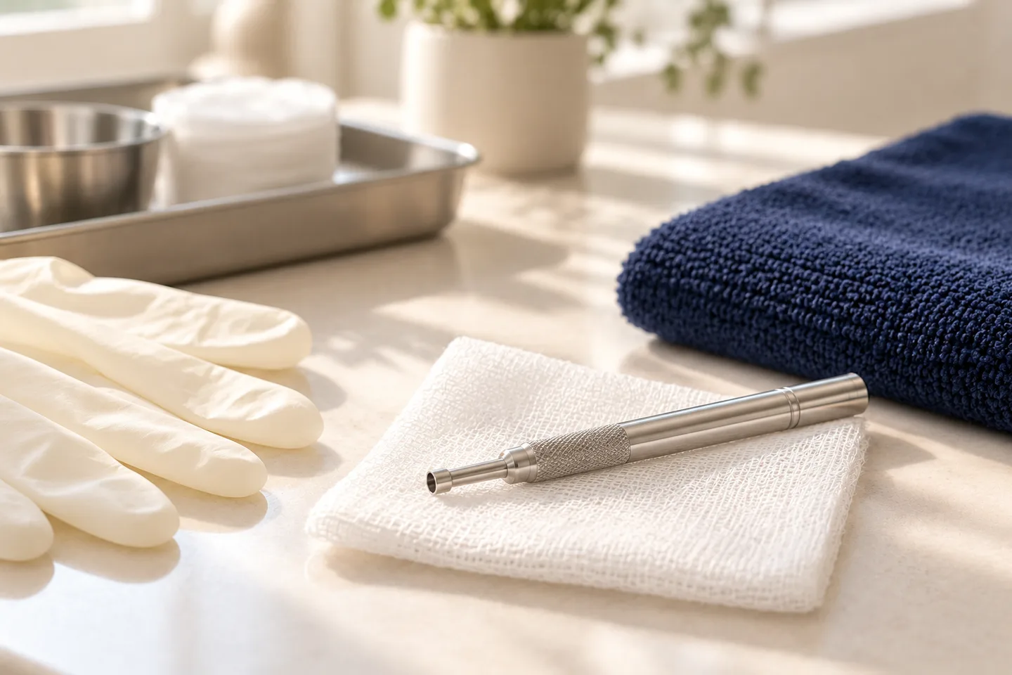
Punch instruments are available in different diameters. The size chosen depends on the suspected diagnosis, anatomical location, and amount of tissue required. Commonly used punches are in the approximately 2–6 mm range, with 3–4 mm sizes frequently used.
The goal is not simply to “take a piece of skin.
” A useful biopsy requires the dermatologist to select:
- the right lesion
- the right part of that lesion
- the appropriate depth
- the appropriate biopsy technique
- the correct laboratory test and specimen handling
These decisions can significantly affect how useful the pathology result will be.
Why Would a Dermatologist Recommend a Skin Biopsy?
Many dermatologic conditions can look similar on the surface. A red scaly patch, for example, might represent an inflammatory disorder, infection, precancerous change, or skin cancer.
A biopsy allows the tissue architecture and individual cells to be examined microscopically. This process is called histopathology.
A dermatologist may recommend a biopsy when a lesion is clinically suspicious, when several diagnoses remain possible, when a persistent skin condition has not behaved as expected, or when knowing the exact diagnosis would change treatment. Skin biopsy is also an essential diagnostic method when skin cancer is suspected.
Importantly, a biopsy is a diagnostic procedure rather than a diagnosis itself. The result needs to be interpreted together with the appearance, location, symptoms, duration, previous treatment, and other clinical findings.
What Conditions Can a Punch Biopsy Help Diagnose?
1. Inflammatory Skin Diseases
Punch biopsy is particularly useful for many inflammatory disorders because it provides tissue from multiple layers of the skin.
Depending on the clinical situation, biopsy may help evaluate conditions such as:
- psoriasis
- lichen planus
- lupus and other connective-tissue diseases
- vasculitis
- certain types of dermatitis
- granulomatous disorders
- blistering diseases
The timing and location of the biopsy matter. For example, a recently developed lesion may be preferable for some inflammatory diseases, while certain blistering disorders require a sample from skin next to the blister for direct immunofluorescence (DIF).
This is one reason the most visibly damaged part of a rash is not necessarily the best place to biopsy.
2. Persistent or Unexplained Rashes
A biopsy may be considered when a rash persists despite treatment, has an unusual appearance, or cannot be confidently diagnosed from examination alone.
However, biopsy does not automatically provide a single definitive answer for every rash. Different inflammatory diseases can produce overlapping microscopic patterns, so correlation between the clinical examination and pathology findings is important.
3. Suspected Skin Cancer
A skin biopsy is required to establish a definite diagnosis of skin cancer. The tissue is examined microscopically to determine whether cancer cells are present and, when relevant, the type and other pathological characteristics.
A punch biopsy may be appropriate for selected lesions where conditions such as basal cell carcinoma (BCC) or squamous cell carcinoma (SCC) are suspected.
But this does not mean every suspicious lesion should undergo punch biopsy.
What about melanoma?
When melanoma is suspected, the choice of biopsy technique requires particular care.
When feasible, complete removal of the suspicious lesion by excision biopsy is generally preferred because a partial sample can miss a diagnostically important area within a heterogeneous pigmented lesion. There are situations—such as unusually large lesions or difficult anatomical sites—where a partial biopsy may be considered, but this should be planned appropriately.
This distinction is important: punch biopsy is useful, but it is not automatically the best biopsy for every suspicious mole.
4. Skin Infections
Selected persistent, deep, or unusual infections may require tissue sampling.
When deep fungal or atypical mycobacterial infection is suspected, specimen handling may differ from an ordinary biopsy. Tissue may need to be divided so that appropriate samples can be sent for histopathology and microbiological testing.
5. Hair and Scalp Disorders
Punch biopsy can sometimes help distinguish different forms of hair loss, particularly when a dermatologist needs to determine whether follicles are being permanently damaged or when the clinical diagnosis remains uncertain.
The biopsy site and the way the specimen is processed are important because the pathologist needs an appropriate view of the hair follicles.
Punch Biopsy vs Shave Biopsy vs Excision Biopsy
There is no single biopsy method that is best for every skin lesion.
Biopsy
type
What is removed Common reason for choosing it
Punch
biopsy
Small cylindrical full-thickness sample
Inflammatory diseases, selected tumors, conditions requiring assessment of deeper skin
Shave
biopsy
Superficial portion of a lesion
Selected superficial or raised lesions
Incisional
biopsy
Part of a larger lesion Larger or deeper processes when representative tissue is required
Excision
biopsy
Entire lesion, usually with a margin
Selected tumors and suspicious pigmented lesions, including suspected melanoma when feasible
A superficial shave sample does not provide the same depth as a punch biopsy and can therefore be unsuitable when the depth of a disease process is important. Conversely, taking a deep punch is unnecessary for some superficial lesions.
The diagnosis should determine the biopsy technique—not the other way around.
How Is a Punch Biopsy Performed?
Step 1: Dermatology Assessment
Before performing the biopsy, the dermatologist examines the lesion and reviews relevant history.
You may be asked about:
- when the lesion appeared
- whether it has changed
- itching, pain or bleeding
- previous treatment
- previous skin cancer
- medications
- allergies
- bleeding disorders
- medications that affect bleeding
- relevant medical conditions
For pigmented lesions, examination may also include dermoscopy, which allows structures beneath the skin surface to be assessed under magnification.
Step 2: Selecting the Biopsy Site
This is one of the most important parts of the procedure.
For inflammatory skin disease, sampling an old, scratched, infected, or heavily treated area may produce nonspecific findings. For other diseases, the active edge rather than the center may contain the most useful diagnostic information.
Occasionally, more than one biopsy is required.
Step 3: Local Anesthesia
The skin is cleaned and local anesthetic is injected around the biopsy site.
The injection can briefly sting, but once the area is numb, patients should generally feel pressure or movement rather than sharp pain during tissue removal. Punch biopsy is ordinarily performed under local anesthesia.
Step 4: Removing the Tissue Sample
The dermatologist places the circular punch perpendicular to the skin and gently rotates it through the required skin layers.
The small cylindrical specimen is then carefully removed so that the tissue is not damaged before microscopic examination.
Step 5: Closing the Wound
Depending on the punch size, location and wound, the site may be closed with one or more stitches. Very small wounds may sometimes be allowed to heal without suturing.
A dressing is then applied.
Step 6: Sending the Sample for Testing
For routine histopathology, the specimen is generally preserved in formalin and sent to a pathology laboratory.
The tissue is processed into very thin sections, stained—most commonly initially with hematoxylin and eosin (H&E)—and examined microscopically by a pathologist or dermatopathologist. Additional stains or tests may sometimes be required.
Does a Punch Biopsy Hurt?
Most patients tolerate punch biopsy well.
The part patients commonly notice most is the brief sting or burning sensation from the local anesthetic injection. Once the area is numb, the actual biopsy should not normally cause sharp pain.
Mild soreness may occur after the anesthetic wears off.
Pain that becomes progressively worse rather than improving—especially when accompanied by redness, warmth, swelling, discharge or fever—should be medically assessed.
Will a Punch Biopsy Leave a Scar?
Yes, some degree of scarring is possible whenever skin is cut.
A punch biopsy usually produces a small scar, but its final appearance depends on factors including:
- biopsy diameter and depth
- anatomical location
- wound tension
- infection or other healing complications
- individual healing tendency
- tendency to form hypertrophic or keloid scars
Skin biopsy can occasionally result in a raised or keloid scar, with some anatomical areas more prone to this type of healing.
For cosmetically sensitive areas such as the face, scar location and biopsy technique should be considered before the procedure.
Recovery and Aftercare
Your dermatologist’s specific wound-care instructions should take priority because care varies depending on the biopsy location and how the wound was closed.
General care commonly involves keeping the wound clean and protected while it heals. Careful wound hygiene following skin biopsy is recommended to reduce complications and support healing.
Avoid repeatedly picking at the wound or scab.
If stitches were placed, you will be told when they should be removed. The timing depends on the anatomical location, wound tension, and individual healing—not one universal number of days for every biopsy.
Contact your doctor if you develop:
- increasing rather than improving pain
- spreading redness
- increasing swelling or warmth
- pus or abnormal discharge
- persistent bleeding
- wound separation
- fever or other signs of infection
What Are the Risks of Punch Biopsy?
Skin biopsy is generally a minor procedure, but it is not risk-free.
Potential complications include:
Bleeding and bruising. Some bleeding can occur during or after the procedure. The dermatologist should know if you take anticoagulants, antiplatelet drugs, or other medications that may affect bleeding.
Infection. Infection is uncommon but possible. Increasing redness, pain, swelling or discharge should be evaluated.
Scarring. A permanent scar can remain at the biopsy site.
Pigmentary change. The healed area may temporarily or permanently appear lighter or darker than the surrounding skin.
Altered sensation. Numbness or unusual sensation can occasionally occur depending on biopsy depth and location.
How Accurate Is a Punch Biopsy?
A well-planned biopsy can provide highly valuable diagnostic information, but no biopsy should be described as 100% accurate.
Its usefulness depends on several factors:
- whether the correct lesion was selected
- whether the correct part of the lesion was sampled
- whether sufficient depth and tissue were obtained
- whether the correct biopsy technique was chosen
- specimen handling and laboratory processing
- correlation between pathology findings and the clinical presentation
Dermatopathology can be complex because different diseases may share similar microscopic patterns. Clinical information supplied to the pathologist—including the lesion’s appearance, location, duration and suspected diagnoses—can therefore be important to interpretation.
Occasionally, a report may be inconclusive and another biopsy or additional testing may be necessary.
What Will the Pathology Report Tell Me?
The answer depends on why the biopsy was performed.
A pathology report may identify:
- a benign skin lesion
- inflammatory skin disease
- infection
- precancerous changes
- basal cell carcinoma
- squamous cell carcinoma
- melanoma or another tumor
- a microscopic pattern that needs correlation with the clinical findings
Additional tests, special stains, immunohistochemistry, microbiological testing, or further tissue sampling may sometimes be required.
A pathology report should therefore be interpreted in context rather than simply viewed as a laboratory result in isolation.
Does a Punch Biopsy Remove the Whole Lesion?
Usually, no.
This is an important distinction for patients.
A punch biopsy is commonly performed to obtain representative tissue for diagnosis, not necessarily to completely remove the lesion.
If the pathology shows a skin cancer or another condition requiring complete removal, additional surgery or treatment may be necessary.
For suspected skin cancer in particular, attempting to completely remove a cancer using a routine punch biopsy is generally not the goal.
Occasionally, a very small lesion may happen to be entirely contained within the punch specimen, but this should not be assumed unless the clinical and pathology findings confirm it.
What If the Biopsy Shows Skin Cancer?
A positive biopsy is the beginning of treatment planning—not the end of the process.
The next step depends on:
- type of skin cancer
- subtype and pathological features
- location
- size
- depth
- whether the lesion was completely sampled or removed
- patient factors
- other findings in the pathology report
Treatment might involve standard surgical excision, Mohs surgery in selected situations, or other approaches depending on the diagnosis.
For melanoma, pathological information such as tumor depth is particularly important for subsequent management.
When Should a Skin Lesion Be Evaluated by a Dermatologist?
Arrange an assessment if you notice a lesion that:
- is new and continues to grow
- changes in size, shape or color
- has multiple or changing colors
- repeatedly bleeds or crusts
- does not heal
- develops persistent ulceration
- becomes painful without an obvious explanation
- looks noticeably different from your other moles
- continues changing over time
Not every changing lesion is cancer, and many benign conditions can look concerning. Conversely, skin cancer does not always look dramatic.
A dermatologist can first determine whether a biopsy is necessary and, if so, which biopsy method is most appropriate.
What to Expect at a Punch Biopsy Consultation in Bangkok
For someone seeking evaluation in Bangkok, the consultation should ideally begin with diagnosis rather than automatically proceeding to biopsy.
A dermatologist may first:
- review your symptoms and medical history
- examine the lesion and surrounding skin
- use dermoscopy when appropriate
- discuss the likely diagnosis and alternatives
- explain whether biopsy is necessary
- select punch, shave, incisional or excisional biopsy according to the clinical question
- explain expected scarring and potential complications
- discuss how the pathology result will affect the next step
International patients should also consider follow-up logistics. If you will leave Thailand shortly after the procedure, ask how pathology results will be communicated, whether stitches need removal, and what would happen if the result requires additional treatment.
That planning is especially important when biopsy is being performed for a suspicious lesion.
Frequently asked questions
Is punch biopsy the same as skin biopsy?
Punch biopsy is one type of skin biopsy. Other techniques include shave, incisional and excisional biopsy.
How big is a punch biopsy?
Punch instruments come in different sizes. Common dermatologic punches are only a few millimeters in diameter, and the appropriate size depends on the lesion, location and diagnostic question.
Do I need stitches after a punch biopsy?
Sometimes. Smaller biopsy wounds may heal without sutures, while others are closed with stitches.
Can a punch biopsy diagnose skin cancer?
Yes. Punch biopsy can diagnose selected skin cancers, but it is not the correct technique for every suspicious lesion.
Is punch biopsy appropriate for melanoma?
Not routinely for a small lesion that can be completely removed. When melanoma is suspected and complete removal is practical, excision biopsy is generally preferred. Partial biopsy may be appropriate in selected situations.
Can a biopsy make cancer spread?
A routine skin biopsy is a standard part of diagnosing suspected skin cancer. There is no clinical reason to avoid an indicated skin biopsy because of a fear that cutting the lesion will make ordinary skin cancer spread.
Can a punch biopsy diagnose a rash?
Yes, particularly when inflammatory skin disease is suspected. However, pathology findings sometimes overlap between conditions, so the result needs to be correlated with the clinical examination.
Can a punch biopsy diagnose fungal infection?
It can contribute to diagnosing selected deep or difficult fungal infections. The dermatologist may need tissue for both histopathology and microbiological testing, which requires appropriate specimen handling.
How long does a punch biopsy take?
The tissue-removal portion is generally brief, but the complete appointment also includes assessment, consent, local anesthesia, bleeding control, possible suturing and dressing.
How long do pathology results take in Bangkok?
Turnaround varies by laboratory, diagnosis and whether special stains, immunohistochemistry or additional testing are required. Patients should be given an expected timeframe by the treating clinic rather than relying on a universal number of days.
Can I exercise after a punch biopsy?
It depends on the location and size of the wound. Activities that stretch, rub or place tension on the biopsy site may need to be temporarily limited. Follow the instructions provided for your specific wound.
Can I shower after a skin biopsy?
Wound-care recommendations depend on the dressing and closure method. Follow the instructions given after the procedure rather than applying a universal rule to every biopsy.
Will I need another procedure if the result is cancer?
Possibly. A punch biopsy often samples only part of a lesion. If cancer is diagnosed, definitive treatment may require further surgery or another treatment chosen according to the cancer type and pathology findings.
Related conditions
-
Skin Biopsy (การวินิจฉัย / Diagnosis) Skin Biopsy (การวินิจฉัย / Diagnosis) A skin biopsy is a minor medical procedure in which a dermatologist removes a small sample of skin so it can be examined under a microscope. It is one of the most important diagnostic tools in dermatology because many skin conditions cannot be confirmed by appearance alone.
-
Shave Biopsy Shave Biopsy A shave skin biopsy is a common dermatology procedure used to remove a superficial portion of a skin lesion for examination under a microscope. It is usually performed under local anesthesia during an outpatient dermatology visit and often does not require stitches.
-
Excisional Biopsy Excisional Biopsy An excisional biopsy means completely cutting out a skin lesion, usually through the full thickness of the skin. The removed tissue is then examined under a microscope.

-
Histopathology Histopathology Histopathology is the microscopic examination of tissue to identify disease. In dermatology, this usually means examining a piece of skin removed during a skin biopsy or surgical excision.
