Melanoma
Melanoma is a serious type of skin cancer that develops from melanocytes, the cells that produce pigment in the skin. Unlike most basal cell carcinomas and many cutaneous squamous cell carcinomas, melanoma has a greater potential to spread to lymph nodes and other organs if it becomes invasive.
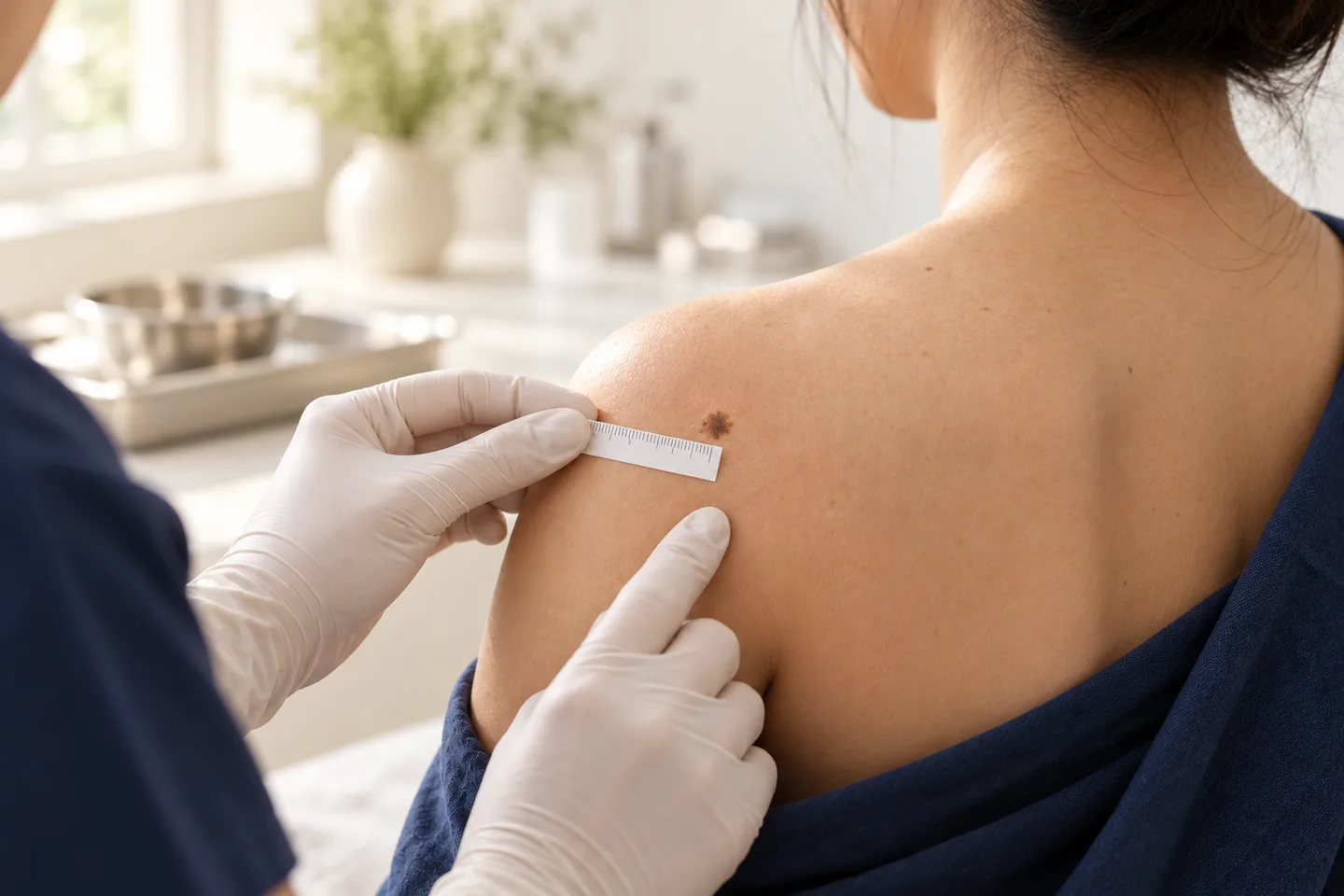
The most important step is early and accurate diagnosis. A suspicious mole does not automatically mean melanoma, but a new, changing, unusual, or bleeding pigmented lesion should be evaluated rather than removed cosmetically without a diagnosis.
For patients diagnosed with melanoma, treatment depends heavily on the depth of the melanoma, ulceration, whether lymph nodes are involved, and whether the cancer has spread elsewhere. Early melanoma is usually treated surgically, while higher-risk or advanced melanoma may require lymph-node assessment, imaging, immunotherapy, targeted therapy, or multidisciplinary cancer care.
For Thai patients, expats, tourists, and international patients seeking melanoma assessment or treatment in Bangkok, obtaining the pathology report and establishing the correct stage are particularly important before deciding on treatment or planning international travel.
What Is Melanoma?
Melanoma is a malignant tumor arising from melanocytes.
Melanocytes produce melanin, the pigment responsible for much of the color in the skin, hair, and eyes.
Melanoma often begins on the skin, but it does not always begin in an existing mole. It can develop as a new lesion on previously normal-looking skin.
It can also occur in areas that receive little sun exposure, including the soles of the feet and beneath the nails. People of all skin colors can develop melanoma.
The major concern with melanoma is its ability to invade deeper into the skin and potentially spread through lymphatic channels or the bloodstream.
This makes early recognition and appropriate biopsy particularly important.
Is Melanoma the Same as Other Skin Cancers?
No.
The three skin cancers patients most commonly encounter are:
- Basal cell carcinoma
- Cutaneous squamous cell carcinoma
- Melanoma
They behave differently.
Basal Cell Carcinoma
Basal cell carcinoma usually grows locally and rarely spreads to distant organs.
Squamous Cell Carcinoma
Cutaneous squamous cell carcinoma can invade surrounding tissue and, particularly when high risk, can spread to lymph nodes or other sites.
Melanoma
Melanoma has a greater potential for regional and distant spread and therefore requires accurate assessment of tumor depth and stage.
This is why melanoma treatment cannot be planned simply from the visible size of a mole.
What Causes Melanoma?
Melanoma develops when melanocytes acquire genetic changes that allow them to grow abnormally.
Ultraviolet radiation is an important preventable risk factor for many cutaneous melanomas, but melanoma is not exclusively a sun-related disease.
It can develop in sun-protected areas as well.
Risk factors may include:
- Significant ultraviolet exposure
- History of severe sunburn
- Fair skin that burns easily
- Numerous moles
- Atypical moles
- Personal history of melanoma
- Family history of melanoma
- Certain inherited genetic conditions
- Reduced immune function
- Increasing age
However, having a risk factor does not mean a person will develop melanoma, and melanoma can occur in people without obvious risk factors.
Can People With Asian or Darker Skin Develop Melanoma?
Yes.
Melanoma can occur in every skin tone.
An important point for Asian patients and people with darker skin is that melanoma may occur on sites that receive relatively little sun exposure, including:
- Palms
- Soles
- Fingers
- Toes
Around or underneath the nails
Clinical guidelines emphasize examining the palms, soles, scalp, nails, and other less-visible areas when checking for melanoma.
A dark streak in a nail is not automatically melanoma, but a new or changing pigmented nail lesion should be evaluated appropriately.
What Does Melanoma Look Like?
Melanoma does not have one single appearance.
It may appear as:
- A new dark spot
- A changing mole
- An irregularly shaped pigmented lesion
- A mole containing several colors
- A black or dark-brown lesion
- A pink or red lesion
- A raised nodule
- A lesion that bleeds
- A lesion that develops a sore or ulcer
- A changing dark streak beneath a nail
Some melanomas contain very little pigment and may appear pink, red, or skin-colored.
Therefore, melanoma should not be thought of only as a “black mole.
”
The ABCDE Warning Signs of Melanoma
The ABCDE rule is a practical way to recognize some suspicious pigmented lesions.
A — Asymmetry
One half of the lesion does not resemble the other half.
B — Border
The border may be irregular, poorly defined, scalloped, or uneven.
C — Color
There may be multiple colors within one lesion, such as:
- Brown
- Black
- Tan
- Red
- White
- Blue
D — Diameter
Melanomas are often larger than 6 mm when diagnosed, but melanoma can be smaller than 6 mm.
A small lesion should not be considered safe based on size alone.
E — Evolving
The lesion changes over time.
Changes may involve:
- Size
- Shape
- Color
- Height
- Surface
- Symptoms
Clinical experts recommend evaluation of lesions showing ABCDE warning signs or lesions that are new, different from others, changing, itching, or bleeding.
Many people have multiple moles that resemble one another.
A lesion that looks noticeably different from the person’s other moles may deserve closer evaluation.
This concept is sometimes called the ugly duckling sign.
For example, a patient may have many small, light-brown moles but develop one new, very dark irregular lesion.
Even if that lesion does not meet every ABCDE criterion, the fact that it looks different can be clinically important.
Can Melanoma Be Smaller Than 6 mm?
Yes.
The “D” in ABCDE is useful, but it should not be interpreted as meaning that lesions smaller than 6 mm cannot be melanoma.
Early melanomas can be small.
A small lesion that is clearly changing or looks different from the patient’s other moles may still need assessment.
Can Melanoma Be Pink or Skin-Colored?
Yes.
This is called amelanotic melanoma when the tumor has little or no obvious dark pigmentation.
It may appear:
- Pink
- Red
- Flesh-colored
- Lightly pigmented
This can make diagnosis more difficult because the lesion may resemble a benign growth or another type of skin cancer.
A persistent, changing, or unusual pink lesion should therefore not be dismissed solely because it is not dark.
What Is Acral Melanoma?
Acral melanoma occurs on areas such as:
- Palms
- Soles
- Fingers
- Toes
- Nail regions
These melanomas deserve particular awareness in Asian and darker-skinned populations because melanoma in these patients may occur in areas not usually associated with heavy sun exposure.
A changing pigmented lesion on the sole should not automatically be assumed to be a bruise, callus, wart, or benign mole.
Can Melanoma Develop Under a Nail?
Yes.
Melanoma can develop in the nail unit.
Warning signs can include:
- A new dark longitudinal band
- A band becoming wider
- Irregular pigmentation
- Multiple colors within the band
- Pigment spreading onto surrounding skin
- Nail distortion
- Bleeding or persistent ulceration
Many dark nail streaks are benign, particularly in people with darker skin.
The goal is not to biopsy every pigmented nail but to identify lesions with suspicious clinical features.
Does Every Changing Mole Mean Melanoma?
No.
Benign moles can change, and many lesions that concern patients turn out not to be melanoma.
Common benign conditions that can resemble melanoma include:
- Ordinary melanocytic nevi
- Atypical nevi
- Seborrheic keratoses
- Solar lentigines
- Blood under a nail
- Benign nail pigmentation
- Angiomas
- Certain inflammatory lesions
However, it is not always possible to distinguish these reliably from melanoma using a photograph or self-examination.
A suspicious lesion should be assessed rather than assumed to be benign.
When Should You See a Dermatologist?
Consider prompt evaluation if you notice:
- A new unusual mole
- A mole that is changing
- Increasing asymmetry
- Irregular borders
- New color variation
- Rapid enlargement
- A lesion that looks different from your other moles
- Bleeding without a clear reason
- Persistent ulceration
- A new dark lesion on the palm or sole
- A changing dark band in a nail
- A pink or red lesion that continues to change
- A previously removed melanoma site developing a new lesion
If you have previously had melanoma, a new suspicious lesion should be taken particularly seriously.
How Do Dermatologists Evaluate a Suspicious Mole?
Evaluation usually begins with a medical history and careful examination.
The dermatologist may ask:
- When did you first notice it?
- Has it changed?
- Has it bled?
- Does it itch or hurt?
- Do you have many moles?
- Have you previously had an atypical mole or melanoma?
- Does melanoma run in your family?
- Has the lesion previously been treated?
The dermatologist may then examine the lesion with a dermatoscope.
What Is Dermoscopy?
Dermoscopy uses a specialized magnifying instrument that allows the dermatologist to see structures and pigment patterns beneath the surface of the skin that are difficult to appreciate with the naked eye.
It can improve the clinical assessment of pigmented lesions and help determine whether a lesion should be monitored or biopsied.
However:
Dermoscopy does not replace histopathology when melanoma is suspected.
A definitive diagnosis requires examination of tissue. Dermatological standards state that a skin biopsy is required to know whether a suspicious lesion is skin cancer.
Melanoma is diagnosed through skin biopsy followed by histopathological examination.
The biopsy technique matters because the pathologist needs enough tissue to evaluate features that determine the next stage of management.
Whenever feasible, a suspicious lesion is generally removed in a way that allows accurate assessment of the melanoma’s depth and other pathological characteristics. The choice of biopsy technique should take into account the lesion’s size, location, and clinical circumstances.
Why Should a Suspicious Mole Not Simply Be Lasered Off?
If melanoma is possible, destroying the lesion with a laser, cautery, freezing, or another destructive cosmetic procedure before obtaining an adequate tissue diagnosis can create problems.
The pathologist may no longer have the complete lesion needed to determine:
- Whether melanoma is present
- The melanoma subtype
- How deeply it has invaded
- Whether ulceration is present
- Other pathological features relevant to staging
A lesion that could represent melanoma should therefore be properly assessed before cosmetic destruction.
What Does the Melanoma Pathology Report Tell You?
A melanoma pathology report contains information that helps determine the next step.
Important findings can include:
- Melanoma subtype
- Whether it is melanoma in situ or invasive melanoma
- Breslow thickness
- Ulceration
- Mitotic activity
- Margin status
- Other microscopic features when relevant
The exact information reported depends on the specimen and clinical situation.
What Is Breslow Thickness?
Breslow thickness measures how deeply an invasive melanoma extends into the skin.
It is measured microscopically in millimeters.
This is fundamentally different from measuring how wide the mole appears on the skin.
For example, a melanoma can look relatively small on the surface but have important vertical invasion.
Breslow thickness is one of the key factors used in melanoma staging and in deciding whether additional procedures, such as sentinel lymph node biopsy, should be discussed.
What Does Ulceration Mean?
Ulceration is a microscopic finding in which the normal epidermal covering over the melanoma is absent due to tumor-related disruption.
It is an important pathological feature used in melanoma staging.
It does not simply mean that the patient scratched the mole.
What Is Melanoma In Situ?
Melanoma in situ, also called stage 0 melanoma, means melanoma cells remain confined to the epidermis, the outermost layer of the skin.
They have not invaded into the deeper dermis.
Surgical excision is the standard treatment for most melanoma in situ.
Melanoma in situ should still be treated. It is not the same as a harmless atypical mole.
What Is Invasive Melanoma?
Invasive melanoma means melanoma cells have extended beyond the epidermis into deeper skin.
Once melanoma becomes invasive, tumor thickness and other pathological features become particularly important because they influence the risk of spread and the treatment plan.
Understanding Melanoma Stages
Melanoma staging is more complex than simply labeling a melanoma as “early” or “advanced.
”
In broad terms:
Stage 0
Melanoma in situ. Cancer cells remain within the epidermis.
Stage I
Invasive melanoma confined to the skin with relatively favorable pathological characteristics.
Stage II
Melanoma remains localized to the skin but has higher-risk characteristics related mainly to tumor thickness and ulceration.
Stage III
Melanoma has spread regionally, such as to nearby lymph nodes or nearby skin or lymphatic sites.
Stage IV
Melanoma has spread to distant organs, distant lymph nodes, or distant skin sites.
The exact stage should be established using the pathology and appropriate clinical investigations rather than estimated from the visible appearance of the mole.Does Every Melanoma Patient Need a CT, MRI, or PET Scan?
No.
Patients with early melanoma generally do not automatically require extensive imaging.
Imaging may be considered when the melanoma’s stage, symptoms, examination findings, or other features create concern about regional or distant spread.
Tests may include:
- Ultrasound
- CT
- MRI
- PET/CT
The appropriate investigation depends on the clinical situation.
Unnecessary imaging can identify incidental abnormalities that are unrelated to melanoma, so testing should be guided by stage and clinical need rather than performed routinely in every patient.
What Is a Sentinel Lymph Node Biopsy?
A sentinel lymph node is the first lymph node, or group of nodes, to which melanoma cells would be expected to travel from the original tumor.
A sentinel lymph node biopsy is a surgical procedure that removes these selected lymph nodes so they can be examined microscopically for melanoma.
The procedure is used for staging rather than simply removing the melanoma from the skin.
Does Every Melanoma Need a Sentinel Lymph Node Biopsy?
No.
Very early, thin melanomas generally do not require sentinel lymph node biopsy.
The decision becomes more relevant as the pathological risk of lymph-node spread increases.
Factors considered can include:
- Breslow thickness
- Ulceration
- Other high-risk pathological features
- Patient factors
The decision should be made after reviewing the complete pathology report.
Why Is Sentinel Lymph Node Biopsy Important?
Its primary role is staging.
If melanoma cells are found in a sentinel lymph node, the melanoma is considered to have regional lymph-node involvement, which changes the stage and may influence further treatment.How Is Early Melanoma Treated?
For localized melanoma, surgery is the cornerstone of treatment.
Treatment usually involves wide local excision.
The melanoma site and a planned margin of normal-looking surrounding skin are removed.
The width of this surgical margin depends largely on the melanoma’s pathological characteristics, particularly Breslow thickness.
There is not one surgical margin appropriate for every melanoma.
Why Is More Skin Removed After the Initial Biopsy?
Patients are sometimes surprised when the original mole appears completely removed by the biopsy but further surgery is still recommended.
The first biopsy primarily establishes the diagnosis and determines important pathological features.
Wide local excision is then performed to remove an appropriate margin around the original melanoma site.
This reduces the chance that microscopic melanoma cells remain around the primary site.
Wide Local Excision for Melanoma
Wide local excision involves removing:
The previous melanoma/biopsy site + a planned margin of surrounding normal-looking skin
The specimen is then evaluated histologically.
Advantages
Standard treatment for localized melanoma
Removes microscopic disease around the primary site
Provides tissue for margin assessment
Often the main treatment needed for early melanoma
Limitations and Possible Complications
Depending on the size and location, surgery may cause:
- Scarring
- Bleeding
- Bruising
- Infection
- Wound separation
- Numbness
- Delayed wound healing
- Need for a skin flap or graft
The size of the final surgical wound can be considerably larger than the original mole.
That does not mean the melanoma itself was necessarily that large.
Does Melanoma Always Need Mohs Surgery?
No.
Conventional wide local excision remains the cornerstone for most cutaneous melanomas.
Mohs micrographic surgery or other staged excision techniques may have a role in selected situations, particularly some forms of melanoma in situ of the lentigo maligna type and anatomically constrained sites where tissue preservation is important.
Mohs surgery should not automatically be assumed to be the best treatment for every melanoma.
What Is Lentigo Maligna?
Lentigo maligna is a form of melanoma in situ that typically develops on chronically sun-damaged skin, often on the head or neck.
Its visible border can sometimes underestimate its microscopic extension.
Surgical management may therefore require careful margin assessment.
Staged excision or Mohs-type approaches can be considered in selected cases, particularly where conserving normal tissue is important.
Can Melanoma In Situ Be Treated With Cream?
Surgery is preferred when feasible.
For lentigo maligna-type melanoma in situ, nonsurgical treatments such as topical imiquimod or radiation may occasionally be considered when surgery is impractical or contraindicated.
However, these approaches generally have lower cure rates than surgery and are not considered equivalent replacements for surgical treatment in routine cases.What Happens If Melanoma Has Spread to Lymph Nodes?
Regional lymph-node involvement generally places melanoma within stage III disease.
Management becomes more complex and may involve:
Surgery
Assessment of the involved lymph-node basin
Imaging
Molecular testing when appropriate
Immunotherapy
Targeted therapy in selected tumors
Multidisciplinary oncology care
Modern management does not automatically require complete removal of an entire lymph-node basin simply because microscopic melanoma is detected in a sentinel node. In appropriate patients, close nodal ultrasound surveillance may be used instead, while complete lymph-node surgery remains relevant in selected situations.What Is Advanced or Metastatic Melanoma?
Advanced melanoma includes disease that cannot be adequately managed with local surgery alone or has spread to distant sites.
Stage IV melanoma can involve organs or distant tissues such as:
- Lung
- Liver
- Brain
- Bone
- Gastrointestinal tract
- Distant lymph nodes
- Distant skin or soft tissue
Treatment has changed substantially with the development of modern immunotherapy and targeted therapy.Immunotherapy for Melanoma
Immunotherapy helps the immune system recognize and attack melanoma cells.
Checkpoint inhibitors used in melanoma treatment can include:
- Pembrolizumab
- Nivolumab
- Ipilimumab
- Nivolumab combined with relatlimab
- Nivolumab combined with ipilimumab in selected settings
These drugs may be used in certain patients with high-risk resected melanoma or unresectable/metastatic disease, depending on the stage and clinical circumstances.
What Are the Limitations of Immunotherapy?
Immunotherapy can produce important and sometimes durable responses, but it does not work for every patient and can cause significant side effects.
Because checkpoint inhibitors activate immune responses, they can cause inflammation in normal organs.
Potential immune-related adverse effects can involve:
- Skin
- Thyroid and other endocrine organs
- Intestines
- Liver
- Lungs
- Kidneys
- Nervous system
- Other organs
Patients receiving these treatments require oncology supervision and appropriate monitoring.
What Is BRAF Testing?
Some melanomas contain a pathogenic alteration in the BRAF gene.
Testing the tumor for a BRAF mutation can help determine whether targeted treatment is an option.
BRAF status is particularly relevant in advanced melanoma and in some higher-risk melanoma treatment decisions.
It is not a test that determines whether an ordinary mole is melanoma.
Targeted Therapy for BRAF-Mutated Melanoma
For appropriate melanomas carrying certain BRAF mutations, treatment can target the BRAF signaling pathway.
Treatment often combines a BRAF inhibitor with a MEK inhibitor.
Examples include:
- Dabrafenib plus trametinib
- Encorafenib plus binimetinib
- Vemurafenib plus cobimetinib
These treatments are used only when appropriate molecular testing demonstrates a relevant BRAF alteration.What Is Adjuvant Treatment?
Adjuvant treatment is additional treatment given after the melanoma has been surgically removed to reduce the risk of recurrence in patients whose melanoma has a sufficiently high risk of returning.
Depending on the stage and tumor characteristics, this may involve:
Anti-PD-1 immunotherapy
BRAF/MEK targeted therapy for selected BRAF-mutated melanoma
Not every melanoma patient needs adjuvant treatment.
What Is Neoadjuvant Treatment?
Neoadjuvant treatment is systemic treatment given before surgery.
It is increasingly relevant in selected patients with resectable, higher-risk melanoma, particularly certain patients with clinically apparent regional disease.
It is not standard treatment for an ordinary early melanoma detected as a suspicious mole.Can Melanoma Come Back After Treatment?
Yes.
Melanoma can recur:
- At or near the original site
- In nearby skin
- In regional lymph nodes
- At distant sites
The likelihood and pattern of recurrence depend strongly on the original melanoma stage and pathological characteristics.
This is why follow-up should be based on the patient’s stage rather than using one schedule for every melanoma patient.
Follow-Up After Melanoma Treatment
Follow-up may include:
- Examination of the surgical site
- Full-body skin examination
- Lymph-node examination
- Education about self-examination
- Imaging in selected higher-stage patients
- Surveillance for treatment-related complications
The frequency and intensity of follow-up depend on melanoma stage, recurrence risk, and the risk of developing another primary melanoma.
Patients who have had melanoma should continue lifelong skin self-examination.Can Someone Who Has Had Melanoma Develop Another
Melanoma?
Yes.
A previous melanoma increases the importance of ongoing skin surveillance.
Follow-up is not limited to checking the original scar.
The dermatologist should also look for new suspicious lesions elsewhere on the body.
What Should You Look for After Melanoma Treatment?
Seek medical review for:
- A new changing mole
- A new unusual pigmented lesion
- A new lump beneath or near the scar
- New pigmentation developing within the scar
- Persistent swelling
- Enlarged lymph nodes
New unexplained symptoms, particularly in patients with higher-stage disease
The significance of symptoms depends heavily on the original melanoma stage.
Recovery After Melanoma Surgery
Recovery depends on:
Location
- Surgical margin
- Size of the resulting wound
- Method of wound closure
- Whether lymph-node surgery was performed
- Patient health and medications
A small melanoma may be closed directly with stitches.
Larger excisions may require:
- More extensive suturing
- A local skin flap
- Skin grafting
- Specialist reconstruction
Will Melanoma Surgery Leave a Scar?
Yes.
Any surgery that cuts through the skin creates a scar.
The appearance depends on:
- Location
- Size
- Depth
- Skin tension
- Reconstruction method
- Individual scar formation
- Wound complications
Cancer clearance should take priority over trying to minimize the initial size of the scar.
Possible Complications of Melanoma Surgery
Potential complications include:
- Pain
- Bleeding
- Bruising
- Infection
- Wound separation
- Delayed healing
- Scarring
- Numbness
- Sensory changes
- Need for additional surgery
Sentinel lymph node biopsy has additional potential complications, including pain, bruising, fluid collection, infection, temporary sensory changes, and less commonly other complications related to lymphatic surgery or mapping agents.Can You Fly After Melanoma Surgery?
This depends on the procedure.
For international patients in Bangkok, consider:
- Wound size
- Risk of bleeding
- Dressing requirements
- Suture-removal date
- Whether a skin graft or flap was used
- Whether sentinel lymph node biopsy was performed
- When the final pathology will be available
- Whether additional treatment might be required
Travel plans should ideally be discussed before surgery rather than after the procedure has already been performed.
Can International Patients Continue Follow-Up in Their Home Country?
Often, yes.
Good documentation is particularly important.
Before leaving Thailand, patients should ideally have access to relevant records such as:
- Original biopsy pathology report
- Surgical pathology report
- Breslow thickness and other staging information
- Margin status
- Sentinel lymph node pathology, if performed
- Imaging reports, if relevant
- Treatment summary
- Wound-care instructions
Follow-up recommendations
For complex melanoma, pathology slides or tissue blocks may occasionally need review by another pathology service.
What Should International Patients Bring to a Melanoma Consultation in Bangkok?
If melanoma has already been diagnosed elsewhere, bring as much original information as possible:
- Pathology report
- Biopsy report
- Previous surgical report
- Photographs taken before biopsy
- Dermoscopy images if available
- Imaging reports
- Current medication list
- Previous skin cancer history
- Oncology treatment records
- Molecular testing results if already performed
A pathology report stating only “melanoma” without the relevant pathological details may not contain enough information to plan definitive treatment.
What to Expect During a Melanoma Consultation
A melanoma consultation may include several separate questions.
1. Is This Lesion Suspicious?
For a lesion that has not yet been biopsied, the dermatologist assesses its clinical and dermoscopic features.
2. Has Melanoma Been Confirmed?
If a biopsy has already been performed, the pathology report should be reviewed.
3. Is It In Situ or Invasive?
This fundamentally changes staging and management.
4. What Is the Breslow Thickness?
For invasive melanoma, depth is a key feature.
5. Is There Ulceration or Another Important Pathological Feature?
These findings can affect staging.
6. Is Further Surgery Required?
Many patients require wide local excision after the diagnostic biopsy.
7. Should Sentinel Lymph Node Biopsy Be Discussed?
This depends on pathological risk.
8. Is Additional Staging Needed?
Not every patient requires imaging or extensive testing.
9. Is Oncology Referral Required?
Higher-stage melanoma may require surgical oncology or medical oncology care.
Questions to Ask After a Melanoma Diagnosis
- Is this melanoma in situ or invasive melanoma?
- What is the Breslow thickness?
- Is ulceration present?
- What melanoma subtype was identified?
- Are the biopsy margins involved?
- How wide should the definitive excision be?
- Do I need sentinel lymph node biopsy?
- Do I need imaging?
- What stage is my melanoma?
- Should my pathology be reviewed?
- Do I need molecular testing such as BRAF testing?
- Do I need an oncologist?
- Do I need adjuvant treatment?
- How should I check my skin after treatment?
- How often should I have professional skin examinations?
Frequently asked questions
Is melanoma a skin cancer?
Yes. Melanoma is a malignant tumor arising from melanocytes.
Is melanoma dangerous?
It can be. Its potential to invade and spread makes early diagnosis particularly important.
Does melanoma always start from a mole?
No. It can develop in an existing mole or arise as a completely new lesion.
Is every black mole melanoma?
No. Most pigmented lesions are not melanoma.
Can melanoma be brown?
Yes.
Can melanoma be pink?
Yes. Some melanomas have little obvious pigment.
Can melanoma be smaller than 6 mm?
Yes. Small melanomas occur.
Does melanoma hurt?
Often it does not. Pain is not required for melanoma.
Can melanoma itch?
It can, but itching is not specific to melanoma.
Can melanoma bleed?
Yes, particularly as it progresses, although benign lesions can also bleed after trauma.
Can melanoma occur on the sole?
Yes.
Can melanoma occur under a nail?
Yes.
Can Asian patients develop melanoma?
Yes. Melanoma occurs in all skin colors.
Can people with dark skin develop melanoma?
Yes.
What is the ABCDE rule?
It refers to asymmetry, border irregularity, color variation, diameter, and evolution or change.
What is the ugly duckling sign?
It describes a lesion that looks noticeably different from the patient's other moles.
Can a dermatologist diagnose melanoma just by looking?
Clinical and dermoscopic examination can identify suspicious lesions, but tissue examination is required for definitive diagnosis.
Can dermoscopy confirm melanoma?
No. It helps determine suspicion but does not replace histopathology.
Do I need a biopsy?
A suspicious lesion generally requires an appropriate biopsy to establish the diagnosis.
Should a suspicious mole be removed with a laser?
Not before melanoma has been appropriately excluded. Destructive treatment can compromise diagnostic tissue.
What is melanoma in situ?
It is melanoma confined to the epidermis without invasion into deeper skin.
What is invasive melanoma?
It means melanoma cells have invaded beneath the epidermis.
What is Breslow thickness?
It is the microscopic measurement of how deeply invasive melanoma extends into the skin.
Why is Breslow thickness important?
It helps determine stage, surgical management, and whether lymph-node assessment should be considered.
What is ulceration?
It is a pathological feature involving loss of the normal epidermal surface over the melanoma and contributes to staging.
What is the main treatment for early melanoma?
Surgical excision.
Why do I need another operation if the biopsy removed the mole?
The diagnostic biopsy establishes the diagnosis. Wide local excision removes an appropriate margin around the melanoma site.
Does every melanoma need Mohs surgery?
No. Wide local excision is standard for most melanomas.
Can Mohs surgery be used for melanoma?
It can be considered in selected situations, particularly some melanoma in situ of the lentigo maligna type.
What is a sentinel lymph node biopsy?
It is a procedure that removes the first draining lymph node or nodes to check microscopically for melanoma spread.
Does every melanoma need sentinel lymph node biopsy?
No. It is considered based on the melanoma's pathological risk.
Does every melanoma patient need a PET or CT scan?
No. Imaging is guided by stage and clinical circumstances.
What happens if melanoma reaches a lymph node?
It changes the stage and may lead to additional staging and treatment.
What is stage IV melanoma?
It means melanoma has spread to distant sites in the body.
Can advanced melanoma be treated?
Yes. Treatment options include modern immunotherapies, targeted therapies in appropriate tumors, surgery in selected circumstances, and other treatments.
What is immunotherapy?
It is systemic treatment that helps the immune system recognize and attack melanoma.
What is BRAF testing?
It checks the melanoma for certain BRAF gene alterations that can make targeted therapy an option.
Can melanoma come back?
Yes. Recurrence can be local, regional, or distant.
Can I develop another melanoma?
Yes, which is why ongoing skin surveillance is important.
Do I need lifelong skin checks?
Follow-up schedules vary by stage, but patients who have had melanoma should maintain lifelong awareness and self-examination of their skin.
Can tourists be evaluated for melanoma in Bangkok?
Yes, but suspected or confirmed melanoma requires careful planning around biopsy, pathology, definitive surgery, staging when needed, and follow-up.
Can I return home after melanoma surgery?
Often yes, but travel should be coordinated with wound care, pathology results, suture removal, and any additional treatment that may be required.
Related conditions
-
Suspicious Mole Assessment Suspicious Mole Assessment A suspicious mole assessment is a focused dermatology evaluation of a mole or pigmented skin lesion that is new, changing, unusual, bleeding, itching, or simply looks different from your other moles.
-
Dermoscopy Dermoscopy Dermoscopy is a non-invasive examination technique that allows a dermatologist to inspect skin lesions with greater detail than is possible with the naked eye alone.

-
Full Body Skin Examination Full Body Skin Examination A full body skin examination is a head-to-toe assessment of the skin performed to look for suspicious moles, abnormal growths, precancerous changes and signs of skin cancer.

-
Skin Biopsy (การวินิจฉัย / Diagnosis) Skin Biopsy (การวินิจฉัย / Diagnosis) A skin biopsy is a minor medical procedure in which a dermatologist removes a small sample of skin so it can be examined under a microscope. It is one of the most important diagnostic tools in dermatology because many skin conditions cannot be confirmed by appearance alone.