Margin Assessment
Margin assessment is one of the most important parts of skin cancer surgery.
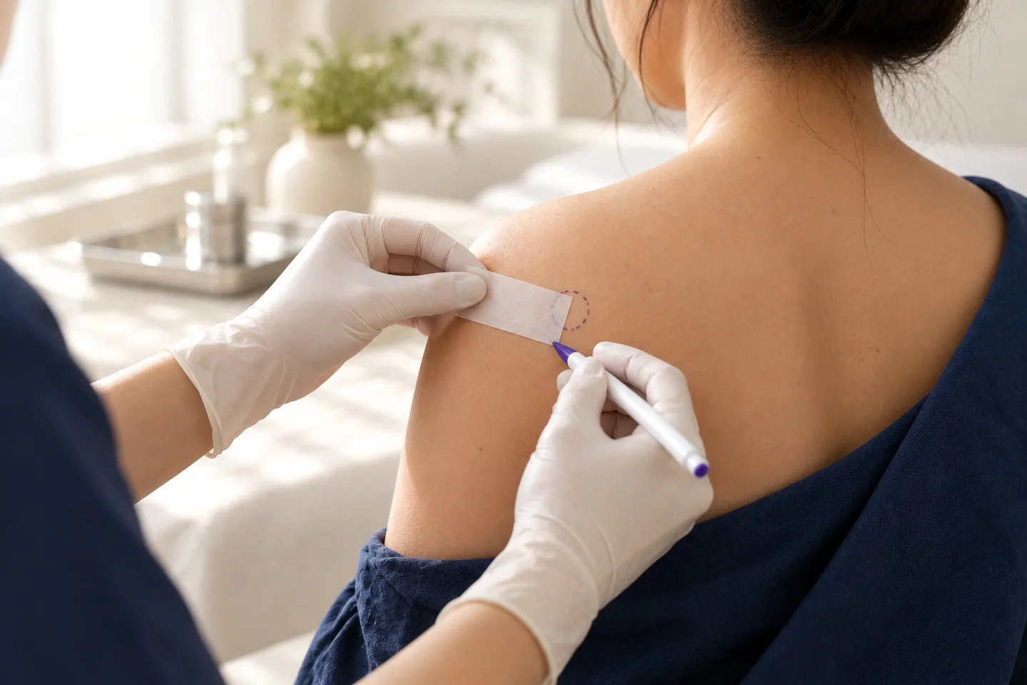
When a skin cancer is surgically removed, the visible tumor is not the only tissue that matters. Cancer cells can sometimes extend microscopically beyond what the doctor can see. For this reason, surgery usually removes the tumor together with a planned amount of surrounding tissue. The removed specimen is then examined by a pathologist to determine whether cancer cells reach the edges of the specimen.
These edges are called surgical margins.
For patients, the pathology report may use terms such as:
- Clear margin
- Negative margin
- Uninvolved margin
- Positive margin
- Involved margin
- Close margin
- Peripheral margin
- Deep margin
Understanding these terms is important because the result can determine whether treatment is complete or whether further surgery or another treatment should be considered.
For patients undergoing skin cancer surgery in Bangkok, especially tourists and international patients, the final pathology and margin assessment should be incorporated into the follow-up plan before care is considered complete.
What Is Margin Assessment in Skin Cancer Surgery?
Margin assessment is the microscopic examination of the edges of tissue removed during skin cancer surgery.
The purpose is to answer a clinically important question:
Does the cancer extend to the edge of the removed specimen?
If cancer cells are present at the edge, some tumor may remain in the patient’s skin.
If the examined margins are free of cancer, the pathology may report the margins as clear or negative.
Surgical excision with margin evaluation is a standard treatment option for localized basal cell carcinoma and cutaneous squamous cell carcinoma.
Margin assessment is also important in melanoma surgery, although melanoma uses different principles for determining the appropriate clinical excision margin.
Why Are Surgical Margins Important?
A skin cancer does not always end exactly where its visible border ends.
Microscopic extensions of cancer may exist beyond the portion seen with the naked eye or dermoscopy.
This is particularly important in tumors that:
- Have poorly defined borders
- Have aggressive histological subtypes
- Have previously been treated
- Have recurred
- Grow along nerves
- Are located in anatomically high-risk areas
- Extend deeper than expected
- The surgeon therefore removes an additional amount of apparently normal skin around the lesion.
- This is called the clinical surgical margin.
- After removal, the pathologist examines the specimen to assess the histological margin.
- These are related concepts, but they are not the same measurement.
Clinical Margin vs Histological Margin
Patients sometimes become confused because their surgeon may say, for example, that a certain clinical margin was taken, but the pathology report later gives a different microscopic distance.
This is normal.
Clinical Surgical Margin
The clinical margin is the amount of normal-looking tissue the surgeon plans to remove around the visible tumor or biopsy site.
It is measured on the patient before the tissue is removed.
Histological Margin
The histological margin is the microscopic distance between the cancer and the edge of the removed specimen.
It is measured by the pathologist after surgery.
These numbers should not be expected to be identical.
Tissue can change shape after removal, fixation and processing, and the microscopic measurement represents the relationship of the tumor to the actual specimen edge rather than simply reproducing the surgeon’s skin marking.
What Are Peripheral and Deep Margins?
Skin cancer specimens have different types of margins.
Peripheral Margin
The peripheral margin, sometimes called the lateral margin, refers to the outer edge around the sides of the tumor.
Imagine looking down at the skin from above: the peripheral margin surrounds the lesion.
Deep Margin
The deep margin is the tissue underneath the tumor.
For an invasive cancer, the surgeon needs to remove the tumor not only sideways but also to an appropriate depth.
A pathology report may therefore state:
- Peripheral margins clear
- Deep margin clear
- Tumor present at peripheral margin
- Tumor present at deep margin
- The location of an involved margin can influence the next surgical plan.
What Does a Clear Margin Mean?
A clear margin, also called a negative or uninvolved margin, means the pathologist does not identify cancer cells at the examined edge of the specimen.
This generally supports that the tumor has been removed at the assessed margins.
However, a clear margin does not mean that recurrence is mathematically impossible.
The interpretation still depends on:
- Type of skin cancer
- Tumor risk
- Histological subtype
- Method used to assess the margins
- Whether the tumor was recurrent
- Other high-risk pathology findings
Conventional excision pathology examines representative sections of the specimen rather than evaluating every microscopic point along the entire edge. Sampling can therefore create some degree of uncertainty even when margins are reported as tumor-free.
Does a Clear Margin Mean the Skin Cancer Is Cured?
A clear margin is reassuring, but it should not be described as a guarantee that the cancer can never recur.
It means no cancer was identified at the margins that were examined.
Recurrence risk is influenced by many other factors, including tumor biology and the method of margin assessment.
In addition, patients who have developed one skin cancer can develop an entirely new skin cancer in another location.
Follow-up therefore remains important.
What Does a Positive Margin Mean?
A positive margin means that cancer cells extend to the edge of the tissue removed.
Other terms that may mean the same thing include:
- Involved margin
- Tumor present at margin
- Carcinoma extends to margin
- Margin involved by tumor
- A positive margin raises concern that residual cancer remains in the patient.
- Further management may include:
- Re-excision
- Mohs surgery
- Another form of margin-controlled surgery
- Radiation therapy in selected circumstances
- Other treatment depending on the cancer type
Repeat excision may be required when surgical margins are inadequate after conventional excision.
Does a Positive Margin Mean the Cancer Has Spread?
No.
This is an important distinction.
A positive surgical margin means:
Cancer reaches the edge of the removed tissue.
It does not automatically mean that the cancer has spread to lymph nodes or distant organs.
Margin status describes local surgical clearance.
Metastasis is a separate issue.
What Is a Close Margin?
A close margin generally means the tumor does not reach the edge, but the microscopic distance between the tumor and the edge is small.
This is different from a positive margin.
For example:
Positive margin: tumor touches the specimen edge.
Close but negative margin: there is still some cancer-free tissue between the tumor and the edge.
Whether a close margin requires further treatment is not determined by one universal distance.
The decision depends on:
- Type of skin cancer
- Histological subtype
- Tumor risk
- Anatomical location
- Whether the tumor is recurrent
- Which margin is close
- Whether additional surgery would create major functional or cosmetic consequences
For cutaneous squamous cell carcinoma, contemporary pathology guidance recognizes margin status as an important prognostic factor, while the precise prognostic meaning of a particular microscopic distance is not as firmly established across all tumors.
Is a 1 mm Clear Margin Enough?
There is no universal answer.
A 1 mm histological clearance might have different implications for:
- A small low-risk basal cell carcinoma on the trunk
- A recurrent infiltrative basal cell carcinoma on the nose
- A poorly differentiated squamous cell carcinoma
- Melanoma in situ
- Invasive melanoma
The pathology result should therefore not be interpreted using a single rule such as:
“Anything more than 1 mm is safe.
”
Margin interpretation must be diagnosis-specific.
How Are Skin Cancer Margins Examined?
After excision, the specimen is sent to pathology.
The pathology laboratory may:
- Inspect the specimen macroscopically.
- Measure the specimen.
- Ink the surgical margins.
- Cut the specimen into sections.
- Process the tissue.
- Examine selected sections under a microscope.
- Report whether tumor is present at the examined margins.
- In some specimens, different ink colors or orientation markers may be used so the pathologist can identify which anatomical side corresponds to an involved margin.
- This becomes particularly important if further surgery is needed.
Why Is Specimen Orientation Important?
Suppose the pathology report simply says:
“Tumor present at peripheral margin.
”
If the specimen has not been oriented, the surgeon may not know exactly whether the involved margin was superior, inferior, medial or lateral.
If the specimen is appropriately oriented, the pathology can sometimes identify the specific margin involved.
This may allow a more focused re-excision.
Orientation is particularly helpful for:
- Large tumors
- Anatomically important locations
- Tumors with complex margins
- Specimens where targeted re-excision may be required
Does Standard Histopathology Examine 100% of the Surgical Margin?
Usually not.
This is one of the most important concepts for patients to understand.
In conventional surgical excision, pathology typically examines representative sections of the specimen.
This technique is highly useful and is standard practice, but it does not literally examine every microscopic point along the entire peripheral and deep margin.
Many pathology laboratories examine only a fraction of the total tumor margin after conventional excision, meaning a declaration of tumor-free margins is subject to some sampling limitation.
This is one of the major differences between conventional excision and Mohs micrographic surgery.
What Is Bread-Loaf Sectioning?
Routine excision specimens are commonly processed by taking a series of sections through the tumor and surrounding tissue.
This is sometimes described conceptually as bread-loaf sectioning because the specimen is cut into slices.
The pathologist examines selected microscopic sections from those slices.
This method provides important information about:
- Diagnosis
- Tumor architecture
- Depth
- Histological subtype
- Selected peripheral margins
- Deep margin
- Other high-risk pathological features
- Its limitation is that the entire outer surface of the specimen is not continuously examined.
How Is Mohs Margin Assessment Different?
Mohs micrographic surgery uses a different approach.
The tumor is removed in stages, and the surgical margins are examined during the procedure.
If cancer remains at a particular location, another layer is removed specifically from that area.
This process continues until the examined margins are clear.
The major advantages are:
- More comprehensive margin assessment
- Ability to map residual tumor to a specific location
- Preservation of healthy tissue
- Immediate additional removal when cancer is detected
- These advantages make Mohs particularly useful for selected high-risk tumors and locations where preserving tissue is important.
Does Mohs Surgery Examine the Entire Margin?
Mohs surgery is designed to provide a much more comprehensive evaluation of the peripheral and deep surgical margin than conventional vertical-section pathology.
Rather than sampling small representative portions, the tissue is processed in a way intended to examine the surgical boundary systematically.
This allows the surgeon to continue removing tissue only where cancer remains.
It is particularly useful for cancers with irregular microscopic extensions.
Is Mohs Surgery Better Than Standard Margin Assessment?
- Not for every tumor.
- Mohs is particularly helpful when:
- The tumor is high risk
- The tumor has recurred
- Borders are poorly defined
- Tissue preservation is important
- The lesion occurs on a functionally or cosmetically sensitive site
- The pathology subtype has infiltrative growth
- There is concern about extension beyond what is clinically visible
Mohs is commonly considered for aggressive, large, recurrent cancers and tumors in areas with limited tissue such as the eyelid, nose, ear, scalp, genital region, hand or foot.
For squamous cell carcinoma, additional indications can include tumors near the eye or ear, poorly defined borders, recurrence and suspected growth along a nerve or blood vessel.
Many low-risk skin cancers can still be appropriately treated with conventional surgical excision.
Permanent Sections vs Frozen Sections
Pathologists can examine tissue using different processing techniques.
Permanent Section
This is the standard method used for most routine surgical excision specimens.
The tissue undergoes processing before being embedded, cut into very thin sections and stained for microscopic examination.
It generally provides excellent tissue detail.
Frozen Section
The tissue is rapidly frozen and examined during or shortly after surgery.
Frozen sections can provide quicker information but are used selectively.
Mohs surgery also uses frozen tissue sections in many settings, although the way the specimen is mapped and processed is specialized and different from simply asking for a routine frozen section.
Can Margin Assessment Be Wrong?
No medical test is perfect.
Potential limitations can include:
- Sampling limitations
- Tissue distortion
- Processing artifacts
- Difficult tumor borders
- Inflammation around the tumor
- Scar from previous biopsy or treatment
- Complex histological subtypes
- Difficulty distinguishing tumor cells from surrounding structures
- Orientation problems
This is why the pathology result should be interpreted together with the clinical history.
If the clinical findings and pathology do not fit together, further review may occasionally be appropriate.
Why Might the Surgeon Think the Tumor Is Removed but Pathology Shows a Positive Margin?
The surgeon can see and feel the visible tumor, but microscopic tumor extensions cannot always be detected during the operation.
Certain skin cancers can extend beyond their apparent clinical borders.
Examples include tumors with:
- Infiltrative growth
- Poorly defined borders
- Scar-like appearance
- Previous treatment
- Perineural growth
- Recurrent disease
A positive pathology margin therefore does not necessarily mean the surgery was performed incorrectly.
It may reflect microscopic extension that was not visible clinically.
What Happens If the Margin Is Positive?
The next step should be individualized.
The doctor should review:
- Cancer type
- Which margin is involved
- Histological subtype
- Tumor size
- Anatomical site
- Whether the tumor is primary or recurrent
- Patient health
- Previous treatment
- Reconstruction already performed
- Possible options include the following.
Re-Excision
Additional tissue is removed from the previous surgical site.
This is a common approach when residual local disease is suspected and another standard excision can reasonably achieve clearance.
Mohs Surgery
Mohs may be considered when detailed margin control is particularly important.
This can be useful for:
- Recurrent tumors
- High-risk locations
- Poorly defined borders
- Aggressive histological subtypes
- Situations where healthy tissue preservation is important
Radiation Therapy
Radiation may be considered in selected situations when additional surgery is difficult, contraindicated or part of treatment for higher-risk disease.
The appropriate choice depends on the specific cancer.
Should Every Positive Margin Be Re-Excised?
- Not necessarily in exactly the same way.
- Positive margins generally require careful management because residual tumor may remain.
- However, the next treatment can differ significantly depending on whether the diagnosis is:
- Basal cell carcinoma
- Squamous cell carcinoma
- Melanoma
- Another less common skin cancer
- Patient age, medical condition, anatomical location and previous treatment can also influence the plan.
- The pathology report should therefore be reviewed in full rather than responding only to the word “positive.
”
Margin Assessment in Basal Cell Carcinoma
Basal cell carcinoma frequently grows locally and may extend beyond its clinically visible border.
Surgical excision with margin assessment is an established treatment for localized basal cell carcinoma.
The likelihood of unexpected extension can be influenced by:
- Tumor size
- Location
- Histological subtype
- Recurrent disease
- Border definition
Aggressive or poorly defined basal cell carcinomas may be more suitable for Mohs surgery or another technique offering more detailed margin control.
Which Basal Cell Carcinoma Subtypes Can Be More Difficult at the Margins?
Some growth patterns can extend microscopically beyond what is visible.
Examples may include infiltrative or other aggressive histological patterns.
This is one reason why the pathology subtype should be considered when interpreting a narrow or positive margin.
A margin result should never be interpreted solely from the gross appearance of the original lesion.
Margin Assessment in Squamous Cell Carcinoma
Margin status is an important component of squamous cell carcinoma pathology and management.
Conventional surgical excision with margin evaluation is widely used for localized cutaneous squamous cell carcinoma.
Higher-risk tumors may have a greater risk of recurrence or spread.
Important pathological and clinical features can include:
- Greater tumor depth
- Poor differentiation
- Perineural invasion
- Recurrent disease
- High-risk anatomical location
- Large tumor size
- Immunosuppression
Contemporary guidance identifies margin status as an important prognostic and reporting factor in cutaneous squamous cell carcinoma.
What Is Perineural Invasion?
Perineural invasion means tumor cells are found growing around or within a nerve.
This can be an important high-risk finding in squamous cell carcinoma.
A patient with a high-risk skin cancer who develops:
- Pain
- Numbness
- Tingling
- Muscle weakness
- Other neurological symptoms
- should be evaluated carefully.
- Margin management may be more complex when perineural invasion is present.
Margin Assessment in Melanoma
Melanoma requires its own surgical principles.
The initial biopsy is used to establish key pathological information, including whether the lesion is melanoma in situ or invasive melanoma and, when invasive, the Breslow thickness.
Definitive treatment generally involves wide local excision.
The clinical surgical margin is determined according to melanoma thickness and stage rather than using the same margin strategy as basal cell carcinoma or squamous cell carcinoma.
Pathology then determines whether melanoma is present at the final surgical margins.
How Are Melanoma Margins Reported?
For melanoma specimens, pathology reporting can distinguish between:
- Invasive melanoma at the margin
- Melanoma in situ at the margin
- Peripheral margin
- Deep margin
- Distance from melanoma to the margin
Standard melanoma reporting protocols specifically include the status and distance of melanoma from peripheral and deep margins.
Why Does Melanoma In Situ Need Margin Assessment?
Melanoma in situ remains within the epidermis, but it can extend laterally beyond the clinically visible pigmentation.
This is particularly relevant in lentigo maligna-type melanoma in situ, where microscopic extension can be difficult to predict.
Staged excision techniques and Mohs surgery can provide extensive peripheral margin assessment in selected melanoma in situ, particularly lentigo maligna and anatomically constrained locations.
Does the Pathology Report Give the Exact Margin Distance?
Sometimes.
A report may state something such as:
- Tumor 4 mm from peripheral margin
- Tumor 2 mm from deep margin
- Margins negative
- Tumor extends to deep margin
- Whether exact distances are routinely reported depends on the tumor type, specimen and pathology reporting protocol.
For melanoma, standard protocols specifically allow reporting measured distances between the tumor and peripheral or deep margins.
For cutaneous squamous cell carcinoma, guidance notes that margin status is required and that reporting distance to the margin is useful, although the clinical meaning of a specific microscopic distance is not firmly established in all cases.
What If the Pathology Report Says “Margins Cannot Be Assessed”?
Occasionally margin assessment is limited.
Possible reasons include:
- Fragmented specimen
- Tissue removed in multiple pieces
- Significant cautery artifact
- Orientation cannot be determined
- The original lesion was partially sampled
- Specimen handling prevents reliable interpretation
- The next step depends on the diagnosis and why the margin cannot be assessed.
For a confirmed invasive malignancy, an unassessable margin may require more consideration than it would for a benign lesion.
Why Should a Suspicious Skin Cancer Not Be Destroyed Before Diagnosis?
Treatments such as:
- Laser
- Electrocautery
- Cryotherapy
- Other destructive procedures
- may destroy tissue without leaving an intact specimen for appropriate histopathological assessment.
- This can be problematic if the lesion is actually an invasive skin cancer.
- Surgical excision has the advantage that tissue remains available for:
- Confirming the diagnosis
- Assessing histological subtype
- Measuring depth when relevant
- Evaluating margins
- Identifying other high-risk pathological features
- Diagnosis should therefore come before cosmetic destruction when malignancy is a realistic possibility.
Should Reconstruction Be Delayed Until Margins Are Known?
Not in every case.
Many conventional excisions are reconstructed immediately.
For example, the wound may be:
- Sutured directly
- Repaired with a flap
- Covered with a graft
- However, when there is substantial concern about margin clearance, surgical planning may occasionally be modified.
- The approach depends on:
- Tumor type
- Margin-control method
- Location
- Size of reconstruction
- Risk of needing further excision
- One advantage of Mohs surgery is that margin clearance is assessed before final reconstruction during the same treatment episode.
What Happens If a Positive Margin Is Found After a Flap or Graft?
This can make additional surgery more complex, but further treatment remains possible.
The surgeon must determine:
- Which margin is involved
- Whether the specimen was oriented
- Where residual cancer is most likely located
- Whether the flap or graft must be partially revised
- Whether Mohs surgery would be useful
- Whether another treatment is more appropriate
- This illustrates why tumor risk and margin strategy should ideally be considered before reconstruction.
Can Scar Tissue Make Margin Assessment More Difficult?
Yes.
Previous surgery, biopsy, cryotherapy, laser treatment or other procedures can produce inflammation and scar tissue.
A recurrent cancer may also grow irregularly through scar tissue.
This can make:
- Clinical borders less obvious
- Surgery more difficult
- Histological interpretation more complex
- Recurrent skin cancers are therefore often approached differently from previously untreated tumors.
Why Are Recurrent Skin Cancers More Important for Margin Control?
A recurrent tumor has already survived or returned after previous treatment.
Its microscopic borders may be less predictable, particularly in scar tissue.
Recurrent basal cell and squamous cell carcinomas are among situations where Mohs surgery may be especially appropriate.
Patients with a new growth developing in a previous skin cancer scar should therefore seek medical assessment rather than assuming it is a normal scar change.
What Symptoms Could Suggest Recurrent Skin Cancer?
Possible warning signs include:
- A new lump in the scar
- Persistent crusting
- A sore that repeatedly opens
- Bleeding
- New ulceration
- Progressive thickening
- New pigmentation in a melanoma scar
- Persistent pain
- Numbness or tingling
- A growing lesion next to the original scar
- These symptoms do not prove recurrence, but they deserve evaluation.
Do Clear Margins Eliminate the Need for Follow-Up?
No.
Follow-up remains important for two reasons.
First, some tumors can recur despite apparently adequate treatment.
Second, patients who have had one skin cancer may develop another independent skin cancer elsewhere.
Follow-up may include:
- Examination of the surgical scar
- Examination of nearby skin
- Full-body skin examination when appropriate
- Lymph-node assessment in selected higher-risk cancers
- Education about self-examination
The schedule should be individualized according to the original cancer.
Margin Assessment for International Patients in Bangkok
For tourists, expats and international patients, margin assessment creates a practical issue:
The surgery may be finished before the pathology process is finished.
Patients should therefore know:
- When pathology is expected
- Who will review the result
- How the result will be communicated
- Whether they should remain in Bangkok until the result is available
- What happens if margins are positive
- Whether additional surgery could be required
- Who will manage follow-up if they have already left Thailand
- A patient should not assume that the treatment pathway is complete simply because the wound has been stitched.
What Documents Should International Patients Keep?
Useful records include:
- Initial biopsy report
- Final excision pathology report
- Operative report
- Tumor type and subtype
- Margin status
- Exact margin distances when reported
- Information about which margin was involved, if applicable
- Reconstruction details
- Follow-up plan
- Clinical photographs when relevant
- If another doctor needs to continue treatment abroad, these records can make the next decision much easier.
What Should You Ask About Your Pathology Result?
Important questions include:
- What type of skin cancer was removed?
- Are the margins clear?
- Which margins were examined?
- Is the peripheral margin clear?
- Is the deep margin clear?
- How close is the cancer to the margin?
- Is the margin truly positive or only close?
- Does the tumor have any aggressive histological features?
- Was the specimen oriented?
- Do I need additional surgery?
- Should Mohs surgery be considered?
- Do I need imaging or lymph-node evaluation?
- When should I have my next skin examination?
What to Expect During a Margin Assessment Consultation
A consultation after skin cancer surgery should not focus only on the word “margin.
”
The doctor should review the entire pathology report.
Diagnosis
What type of skin cancer was found?
Histological Subtype
Does it have an aggressive growth pattern?
Margin Status
Are the peripheral and deep margins clear?
Margin Distance
If reported, how close is the tumor to the edge?
High-Risk Findings
Are there features such as perineural invasion, poor differentiation or significant depth?
Surgical Site
Would another excision be straightforward or difficult?
Previous Treatment
Is this a primary or recurrent tumor?
Next Step
Is observation appropriate, or should additional surgery or another treatment be considered?
Frequently asked questions
What is margin assessment in skin cancer surgery?
It is microscopic examination of the edges of tissue removed during skin cancer surgery to determine whether cancer reaches the specimen boundary.
What is a surgical margin?
It is the normal-looking tissue removed around and beneath the skin cancer.
Why does a surgeon remove normal skin?
Because microscopic cancer cells can extend beyond the visible edge of a tumor.
What is a clear margin?
It means no cancer is identified at the examined edge of the specimen.
What is a negative margin?
It generally means the same as a clear margin.
What is a positive margin?
It means cancer cells reach the edge of the removed specimen.
Does a positive margin mean metastatic cancer?
No. It indicates incomplete local clearance may be present, not necessarily distant spread.
What is a close margin?
It means the tumor is near the specimen edge but does not reach it.
Does every close margin need another operation?
No. The decision depends on cancer type, risk factors, location and pathology.
Is a 1 mm clear margin safe?
There is no single microscopic distance that can be considered universally adequate for every skin cancer.
What is a peripheral margin?
It is the outer margin around the sides of the tumor.
What is a deep margin?
It is the surgical boundary beneath the tumor.
Why can the clinical margin differ from the pathology margin?
One is measured on the patient before excision; the other is measured microscopically after the tissue has been removed and processed.
Is the entire margin examined after standard excision?
Usually not. Routine histology generally examines representative sections.
Can standard pathology miss tumor between the examined sections?
Sampling limitations are possible, which is one reason margin-control techniques such as Mohs surgery are used for selected tumors.
How does Mohs surgery assess margins?
The tumor is removed in stages and the surgical margin is examined during the procedure. Additional tissue is removed from areas where cancer remains.
Is Mohs surgery always better?
No. It is particularly valuable for selected high-risk, recurrent or anatomically sensitive tumors.
Can basal cell carcinoma have positive margins?
Yes.
What happens if basal cell carcinoma has a positive margin?
Additional excision or Mohs surgery may be considered depending on the tumor and location.
Can squamous cell carcinoma have positive margins?
Yes. Margin status is an important part of squamous cell carcinoma management.
Is a positive squamous cell carcinoma margin important?
Yes. Residual tumor may remain, and the complete pathology should be reviewed to determine further treatment.
Are melanoma margins different?
Yes. Melanoma clinical excision margins are based primarily on melanoma thickness and stage.
Does melanoma pathology report margin distance?
It can. Standard melanoma reporting protocols include reporting peripheral and deep margin status and allow measurement of distance from the tumor to those margins.
Can melanoma in situ have a positive margin?
Yes.
What happens if melanoma in situ reaches the margin?
Further surgical management is generally considered to obtain appropriate microscopic clearance.
Can pathology tell which side is positive?
Sometimes, particularly when the specimen has been appropriately oriented and marked.
What if the specimen was not oriented?
The pathology may be able to identify that a margin is involved but not precisely which anatomical side corresponds to it.
Can a positive margin be treated with another excision?
Often yes, depending on the diagnosis and location.
Can Mohs surgery be used after incomplete excision?
It may be appropriate for selected tumors, particularly recurrent or high-risk basal cell and squamous cell carcinomas.
Does a clear margin guarantee that cancer will never return?
No.
Do I still need skin checks after clear margins?
Yes. Follow-up can detect recurrence and new primary skin cancers.
Should I wait for pathology before flying home?
When practical, reviewing the final pathology before leaving Bangkok can simplify treatment planning. If you need to travel earlier, there should be a clear system for receiving and acting on the result.
What happens if my margin is positive after I return home?
The pathology report and operative records should be reviewed by the doctor continuing your care so that additional treatment can be planned.
Related conditions
-
Surgical Excision Surgical Excision Surgical excision is one of the main treatments for skin cancer. During the procedure, the doctor removes the cancer together with a planned amount of normal-looking skin around it. The removed tissue is then examined under a microscope to determine whether cancer cells extend to the surgical edges, known as the margins.
-
Skin Biopsy (การวินิจฉัย / Diagnosis) Skin Biopsy (การวินิจฉัย / Diagnosis) A skin biopsy is a minor medical procedure in which a dermatologist removes a small sample of skin so it can be examined under a microscope. It is one of the most important diagnostic tools in dermatology because many skin conditions cannot be confirmed by appearance alone.

-
Histopathology Histopathology Histopathology is the microscopic examination of tissue to identify disease. In dermatology, this usually means examining a piece of skin removed during a skin biopsy or surgical excision.

-
Mohs Surgery / Mohs Referral Mohs Surgery / Mohs Referral Mohs surgery, or Mohs micrographic surgery, is a specialized form of skin cancer surgery in which the tumor is removed in stages and the surgical