Seborrheic Keratosis
Seborrheic Keratosis is one of the most common benign skin growths seen by dermatologists. It often appears as a brown, black, or tan spot that looks as though it is "stuck onto the skin.
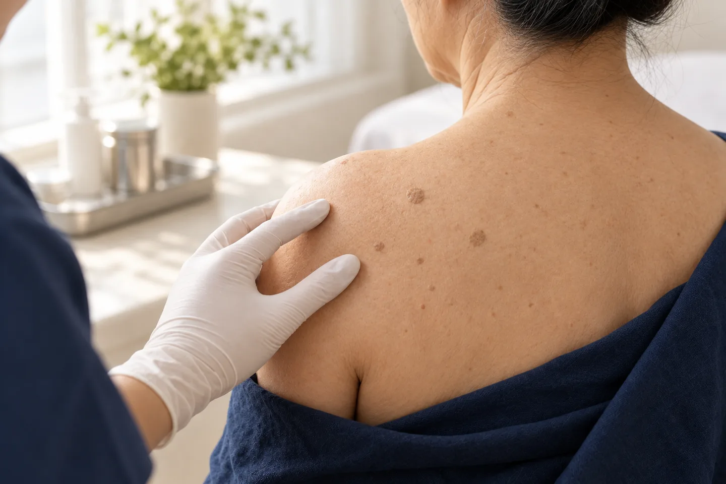
” While these growths are not skin cancer and usually do not require treatment, some people choose to remove them because they become irritated, catch on clothing, or affect their appearance.
The most important step before removal is confirming that the lesion is truly Seborrheic Keratosis and not another condition that may require a different approach, such as melanoma, basal cell carcinoma, or Squamous Cell Carcinoma.
This guide explains how dermatologists diagnose Seborrheic Keratosis, when treatment is appropriate, available removal options, healing expectations, and what international patients visiting Bangkok should know.
What Is Seborrheic Keratosis?
Seborrheic Keratosis is a common benign skin growth that develops from epidermal cells. It is especially common in adults and tends to become more frequent with age.
These growths are not contagious, are not caused by poor hygiene, and do not usually become skin cancer.
Common characteristics
- Brown, black, tan, or skin-colored
- Flat or raised
- Waxy or rough surface
- “Stuck-on” appearance
- Single or multiple lesions
- May slowly increase in number over time
Some people develop only one lesion, while others develop dozens across the face, chest, back, or neck.
5
What Causes Seborrheic Keratosis?
The exact cause is not completely understood, but several factors increase the likelihood of developing these growths.
Risk factors
- Increasing age
- Family history
- Long-term sun exposure (may contribute in some cases)
- Naturally occurring skin aging
Unlike viral warts, Seborrheic Keratosis is not caused by HPV and cannot spread from one person to another.
What Does Seborrheic Keratosis Look Like?
The appearance can vary considerably.
Typical features
- Well-defined edges
- Waxy texture
- Rough or slightly crumbly surface
- Appears attached to the skin surface
- Usually grows slowly
Common locations
- Face
- Neck
- Chest
- Back
- Shoulders
- Abdomen
They are uncommon on the palms and soles.
Is Seborrheic Keratosis Dangerous?
In most cases, no.
Seborrheic Keratosis is considered a benign skin condition.
However, not every brown or raised skin lesion is Seborrheic Keratosis. Some skin cancers can resemble it, especially when a lesion changes or develops unusual features.
A dermatologist should evaluate any lesion that:
- Changes rapidly
- Bleeds repeatedly
- Develops an irregular border
- Has multiple colors
- Forms a persistent sore
- Looks noticeably different from other spots on your body
Conditions That Can Look Like Seborrheic Keratosis
Several skin conditions can appear similar.
Benign conditions
- Melanocytic Nevus
- Skin Tag
- Wart
- Solar Lentigo
- Dermatosis Papulosa Nigra
Conditions requiring careful evaluation
- Melanoma
- Basal Cell Carcinoma
- Squamous Cell Carcinoma
- Bowen’s Disease
- Pigmented Actinic Keratosis
This is why accurate diagnosis should come before cosmetic removal.
How Dermatologists Diagnose Seborrheic Keratosis
Diagnosis usually begins with a clinical examination.
The dermatologist evaluates:
- Color
- Texture
- Borders
- Size
- Growth pattern
- Whether the lesion resembles other spots on your skin
Dermoscopy
4
Dermoscopy allows the dermatologist to examine structures beneath the skin surface that are not visible to the naked eye.
Typical dermoscopic features of Seborrheic Keratosis may include:
- Milia-like cysts
- Comedo-like openings
- Fissures and ridges
- Sharp borders
Dermoscopy often improves diagnostic confidence but does not replace biopsy when the diagnosis remains uncertain.
When Is a Biopsy Needed?
A Skin Biopsy may be recommended if:
- The lesion has atypical features.
- It has recently changed.
- It bleeds repeatedly.
- Melanoma or another skin cancer cannot be confidently excluded.
The tissue can then be examined with Histopathology to establish the diagnosis.
When Should Seborrheic Keratosis Be Removed?
Removal is not medically necessary for most lesions.
Treatment is commonly considered when:
- The lesion catches on clothing.
- It becomes irritated.
- It bleeds after repeated friction.
- It causes cosmetic concerns.
- The diagnosis needs confirmation.
Treatment Options
The best treatment depends on the lesion’s size, thickness, location, and appearance.
Cryotherapy
4
Cryotherapy uses liquid nitrogen to freeze the lesion.
Advantages
- Quick procedure
- No surgical incision
- Suitable for many small lesions
Limitations
- Temporary blistering may occur.
- Pigment changes may develop.
- Thick lesions may require additional treatment.
Cryotherapy is often used for thinner Seborrheic Keratoses.
Shave Removal
4
Shave Removal removes the raised portion of the lesion with a surgical blade after local anesthesia.
Advantages
- Immediate removal
- Usually does not require stitches
- Tissue can be sent for Histopathology when appropriate
Limitations
- Leaves a healing wound
- Color changes or minor scarring may occur
- Not every lesion is suitable for this technique
Curettage
5
Curettage removes the lesion using a specialized instrument that gently scrapes the growth away.
Advantages
- Effective for many raised lesions
- Frequently combined with other techniques
Limitations
- Requires local anesthesia in many cases.
- Healing occurs through the surface wound.
Electrocautery
6
Electrocautery uses electrical energy to remove or destroy the lesion.
Advantages
- Effective for selected lesions
- Often combined with curettage
Limitations
- Temporary redness
- Pigment changes
- Small scars are possible
Laser Treatment
Some lasers may be used for carefully selected benign lesions.
However, laser treatment should not be used simply to remove a pigmented lesion without appropriate evaluation, especially if there is concern about skin cancer.
Which Treatment Is Best?
There is no single best treatment for every patient. Situation
Possible treatment
Small thin lesion Cryotherapy
Raised lesion Shave Removal
Thick waxy lesion Curettage ± Electrocautery
Uncertain diagnosis Biopsy with Histopathology
Cosmetic removal Depends on location and lesion type
The decision should balance cosmetic outcome, healing time, and diagnostic needs.
Recovery After Removal
Healing depends on:
- Treatment method
- Lesion size
- Body location
- Individual healing response
Typical aftercare
- Keep the area clean.
- Follow wound care instructions.
- Avoid picking scabs.
- Protect healing skin from excessive sun exposure.
- Watch for signs of infection.
Some redness during healing is expected.
4
Possible Side Effects
Potential side effects vary by treatment.
They may include
- Redness
- Temporary discomfort
- Blistering (after cryotherapy)
- Scabbing
- Hyperpigmentation
- Hypopigmentation
- Small scars
- Infection (uncommon)
Patients with darker skin tones may have a higher risk of post-inflammatory pigment changes.
Can Seborrheic Keratosis Come Back?
A completely removed lesion usually does not regrow in exactly the same form.
However:
- New Seborrheic Keratoses can develop elsewhere.
- Multiple lesions may continue appearing with age.
- Incompletely treated lesions may persist.
Developing new lesions over time is common and does not necessarily indicate a serious problem.
When Should You See a Dermatologist?
Arrange an evaluation if a skin lesion:
- Changes rapidly
- Bleeds repeatedly
- Becomes painful
- Develops irregular borders
- Has multiple colors
- Looks different from your other spots
- Forms a persistent sore
Early evaluation helps distinguish benign growths from skin cancer.
What to Expect During a Consultation in Bangkok
A dermatology consultation typically includes:
- Medical history.
- Skin examination.
- Dermoscopy when appropriate.
- Discussion of whether removal is recommended.
- Review of treatment options.
- Wound care instructions.
- Histopathology planning if tissue needs examination.
For international patients visiting Bangkok, discussing travel plans beforehand can help with scheduling follow-up care if stitches or pathology results are expected after departure.
Frequently asked questions
Is Seborrheic Keratosis contagious?
No. It cannot spread between people.
Is Seborrheic Keratosis skin cancer?
No. It is a benign skin growth.
Can Seborrheic Keratosis become cancer?
It is generally considered benign, but a lesion that changes or appears unusual should be evaluated because some skin cancers can resemble Seborrheic Keratosis.
Can I remove Seborrheic Keratosis at home?
Home removal is not recommended because other conditions can look similar.
Does removal hurt?
Local anesthesia is commonly used for procedures such as Shave Removal, Curettage, and Electrocautery.
Will it leave a scar?
Any procedure that removes skin tissue can leave some degree of scar or pigment change.
Which treatment has the fastest recovery?
Recovery depends on the lesion and treatment method rather than one universally fastest option.
Can I swim after treatment?
Swimming is usually postponed until the wound has healed sufficiently.
Can I fly after removal?
Many small procedures do not prevent air travel, but wound care and follow-up should be planned in advance.
Will insurance cover removal?
Coverage depends on the reason for treatment and individual insurance policies.
Related conditions
-
Full Body Skin Examination Full Body Skin Examination A full body skin examination is a head-to-toe assessment of the skin performed to look for suspicious moles, abnormal growths, precancerous changes and signs of skin cancer.

-
Dermoscopy Dermoscopy Dermoscopy is a non-invasive examination technique that allows a dermatologist to inspect skin lesions with greater detail than is possible with the naked eye alone.

-
Suspicious Mole Assessment Suspicious Mole Assessment A suspicious mole assessment is a focused dermatology evaluation of a mole or pigmented skin lesion that is new, changing, unusual, bleeding, itching, or simply looks different from your other moles.
-
Skin Biopsy (การวินิจฉัย / Diagnosis) Skin Biopsy (การวินิจฉัย / Diagnosis) A skin biopsy is a minor medical procedure in which a dermatologist removes a small sample of skin so it can be examined under a microscope. It is one of the most important diagnostic tools in dermatology because many skin conditions cannot be confirmed by appearance alone.