






























Bowen’s Disease
Bowen’s disease, also called squamous cell carcinoma in situ, is an early form of squamous cell skin cancer in which abnormal cancerous cells remain confined to the outermost layer of the skin, the epidermis. Because the abnormal cells have not invaded into the deeper skin, Bowen’s disease is considered non-invasive. It is nevertheless a true form of skin cancer and should usually be treated or monitored because a small proportion of lesions can progress to invasive squamous cell carcinoma.
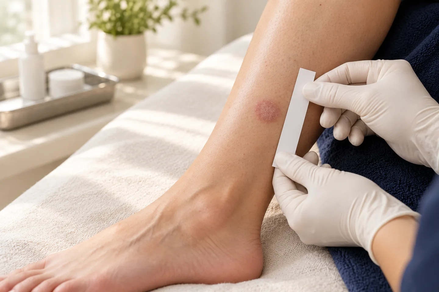
Treatment is not identical for every patient. Depending on the size, thickness, location and number of lesions, options may include surgical excision, curettage and cautery, cryotherapy, topical fluorouracil, topical imiquimod, photodynamic therapy and, in selected circumstances, other approaches. If there is concern that the lesion may already have become invasive, a biopsy or surgical excision is particularly important.
For patients seeking Bowen’s disease treatment in Bangkok, the most important first step is to confirm the diagnosis and determine whether the lesion is truly confined to the epidermis or whether there are clinical features suggesting invasive squamous cell carcinoma.
What Is Bowen’s Disease?
Bowen’s disease is another name for squamous cell carcinoma in situ of the skin.
The term “in situ” means that the abnormal cells remain in the layer where they originated. In Bowen’s disease, abnormal keratinocytes involve the epidermis but have not invaded through the basement membrane into the deeper dermis.
Other terms that may appear on a pathology report include:
- squamous cell carcinoma in situ
- SCC in situ
- intraepidermal squamous cell carcinoma
- intraepidermal carcinoma
- Bowen disease
These terms generally describe the same basic process when referring to the skin.
Is Bowen’s Disease Really Skin Cancer?
Yes.
Bowen’s disease contains malignant squamous cells, so medically it is classified as squamous cell carcinoma in situ.
However, it is important to distinguish it from invasive squamous cell carcinoma.
In Bowen’s disease, abnormal cells are confined to the epidermis. In invasive squamous cell carcinoma, cancer cells have crossed into the deeper layers of the skin and may have access to nerves, blood vessels and lymphatic structures.
That distinction affects both prognosis and treatment options.
Bowen’s Disease vs Invasive Squamous Cell Carcinoma
| Bowen’s Disease | Invasive Squamous Cell Carcinoma |
|---|---|
| Cancer cells remain in the epidermis | Cancer cells have invaded deeper skin |
| Also called squamous cell carcinoma in situ | Also called invasive SCC |
| Usually appears as a slowly growing patch or plaque | May form a thick plaque, nodule, ulcer or rapidly growing lesion |
| Several nonsurgical treatments may be appropriate | Surgery is usually central to treatment |
| No dermal invasion | Dermal invasion is present |
| Lower risk of spread | Some tumors can spread to lymph nodes or beyond |
A lesion cannot always be classified confidently by appearance alone. When invasion is suspected, histopathological examination is important.
What Causes Bowen’s Disease?
The most common cause is long-term ultraviolet damage from sunlight.
Bowen’s disease is particularly associated with chronically sun-exposed skin. Long-term exposure over many years can damage the genetic material inside keratinocytes and eventually contribute to abnormal cell growth.
Risk factors can include:
- long-term sun exposure
- increasing age
- previous skin cancer
- extensive sun damage
- weakened immune function
- organ transplantation
- long-term immunosuppressive medication
- previous radiation exposure to the affected area
- certain chronic skin damage
Some cases may occur in locations that receive relatively little sunlight, so absence of significant sun exposure does not completely exclude the diagnosis.
Does Living in Bangkok Increase the Risk?
Bangkok and Thailand have high levels of ultraviolet exposure throughout much of the year.
However, Bowen’s disease usually reflects cumulative lifetime ultraviolet damage, rather than exposure during only the last few months or years.
A patient who recently moved to Thailand may have accumulated much of their sun damage decades earlier.
For people living long-term in Bangkok, practical sun protection remains important because it may reduce additional ultraviolet damage and the development of further precancerous or cancerous lesions.
What Does Bowen’s Disease Look Like?
The classic appearance is a slowly enlarging red or pink scaly patch or plaque.
It may be:
- red
- pink
- brownish
- dry
- scaly
- crusted
- slightly raised
- sharply or irregularly bordered
- persistent for months or years
Bowen’s disease typically presents as one or more slowly enlarging erythematous, scaly plaques.
Some lesions are thin and relatively flat. Others become thicker, crusted or hyperkeratotic.
Is Bowen’s Disease Usually Painful?
Often it is not.
Some patients have no symptoms other than a persistent patch.
Others may experience:
- itching
- mild tenderness
- irritation
- crusting
- occasional bleeding
Increasing pain or tenderness can be important, especially if accompanied by thickening, ulceration or rapid growth.
Can Bowen’s Disease Look Like Eczema or Psoriasis?
Yes.
This is one of the reasons diagnosis can sometimes be delayed.
Bowen’s disease may resemble:
- eczema
- psoriasis
- fungal infection
- actinic keratosis
- superficial basal cell carcinoma
- seborrheic keratosis
- irritated benign lesions
A useful warning sign is a persistent localized patch that does not behave like an ordinary rash.
For example, a single scaly red patch that remains in approximately the same location for months despite appropriate treatment deserves further evaluation.
Bowen’s Disease vs Actinic Keratosis
Actinic keratosis is a sun-damaged precancerous lesion that may precede squamous cell carcinoma.
Bowen’s disease is further along the disease spectrum because the epidermis contains full-thickness malignant keratinocytes, although there is still no invasion into deeper skin.
Actinic keratosis often feels:
- rough
- gritty
- dry
- scaly
Bowen’s disease more commonly forms a persistent, better-defined scaly plaque.
The two conditions can overlap clinically, and biopsy may sometimes be required to distinguish them.
Bowen’s Disease vs basal cell carcinoma
Bowen’s disease and superficial basal cell carcinoma may sometimes look similar.
Both may present as a red, scaly, slowly growing patch.
However:
Bowen’s disease originates from squamous keratinocytes and represents squamous cell carcinoma in situ.
basal cell carcinoma originates from basal-type cells and has a different biological behavior and treatment pathway.
Clinical examination and dermoscopy may help distinguish them, but biopsy may be necessary when the diagnosis remains uncertain.
Can Bowen’s Disease Turn Into Invasive Squamous Cell Carcinoma?
Yes.
Bowen’s disease is non-invasive at the time of diagnosis, but untreated lesions can occasionally progress into invasive squamous cell carcinoma.
Published sources characterize the risk as relatively low, which is why observation may occasionally be reasonable in selected frail or elderly patients, but dermatologists generally recommend treatment or appropriate monitoring rather than ignoring the lesion.
The most important practical issue is identifying lesions that may already contain an invasive component.
Warning Signs of Possible Invasion
A Bowen’s disease lesion should be reassessed promptly if it develops:
- a new lump or nodule
- increasing thickness
- rapid growth
- ulceration
- persistent bleeding
- marked tenderness or pain
- significant crusting
- firmness beneath the lesion
These features do not prove invasive cancer, but they increase the need for careful assessment and often biopsy.
Where Does Bowen’s Disease Commonly Occur?
It can occur almost anywhere.
Common locations include:
- lower legs
- face
- scalp
- ears
- neck
- arms
- hands
- trunk
Sun-exposed sites are common, but lesions can also occur in covered areas.
Anogenital squamous cell carcinoma in situ requires somewhat different considerations and should be assessed in the context of its anatomical location.
Can You Have More Than One Bowen’s Disease Lesion?
Yes.
Some patients develop multiple lesions, particularly those with extensive sun damage or immune suppression.
When multiple lesions are present, treatment planning may differ from that for a single small lesion.
For example, field-directed or topical treatment may be considered in situations where repeated surgical excision would be impractical.
How Do Dermatologists Diagnose Bowen’s Disease?
Diagnosis begins with the clinical examination.
The dermatologist evaluates:
- location
- size
- surface scale
- thickness
- borders
- color
- ulceration
- growth pattern
- symptoms
- number of lesions
A dermatoscope may be used to examine additional structures within the lesion.
However, a biopsy is often performed when:
- the diagnosis is uncertain
- invasive squamous cell carcinoma is possible
- the lesion is thick
- the lesion is nodular
- there is ulceration
- the lesion is recurrent
treatment planning requires histological confirmation
What Is a Skin Biopsy?
A skin biopsy removes part or all of a suspicious lesion for microscopic examination.
Possible approaches include:
Shave Biopsy
A superficial portion of the lesion is removed.
Punch Biopsy
A small circular sample is taken through several layers of the skin.
Incisional Biopsy
A representative part of a larger lesion is removed.
Excisional Biopsy
The entire lesion is surgically removed.
The technique depends on the lesion’s size, location and degree of suspicion for invasive disease.
What Does the Pathology Report Show?
Typical Bowen’s disease histology demonstrates abnormal keratinocytes throughout the epidermis without invasion through the basement membrane.
A pathology report may contain terms such as:
- squamous cell carcinoma in situ
- Bowen disease
- full-thickness epidermal atypia
- no invasion identified
- involved margin
- clear margin
The exact wording matters because treatment can differ if invasive squamous cell carcinoma is identified.
What Does “No Invasion Identified” Mean?
It means the pathologist did not identify cancer cells extending into the deeper dermis in the tissue examined.
This supports a diagnosis of squamous cell carcinoma in situ rather than invasive squamous cell carcinoma.
However, biopsy only examines the tissue that was sampled. If the clinical lesion is large, thick or irregular and the biopsy sample is small, the dermatologist may still need to consider whether another part of the lesion could contain invasion.
Treatment Options for Bowen’s Disease
Because Bowen’s disease remains confined to the epidermis, several treatment approaches may be reasonable.
The choice depends on:
- lesion size
- lesion thickness
- anatomical location
- number of lesions
- whether invasion is suspected
- previous treatment
- immune status
- wound-healing considerations
- patient preference
Established options include surgical excision, curettage with electrosurgery, cryotherapy, topical fluorouracil, topical imiquimod and photodynamic therapy.
Surgical Excision
Surgical excision removes the entire lesion together with an appropriate border of surrounding skin.
The specimen is then sent for histopathological examination.
Advantages
Excision provides:
- complete tissue for pathological assessment
- information about surgical margins
- the ability to detect unexpected invasive disease
- a straightforward approach for many solitary lesions
Limitations
Potential disadvantages include:
- scar formation
- stitches
- bleeding
- infection
- wound separation
- longer healing in certain locations
- difficulty closing larger defects
Excision is particularly useful when there is concern that the lesion may contain an invasive component.
Does Every Bowen’s Disease Lesion Need Surgery?
No.
Unlike invasive squamous cell carcinoma, Bowen’s disease can often be treated successfully with nonsurgical methods.
Surgery may be particularly attractive when:
- the lesion is solitary
- invasion is suspected
- histological confirmation of complete removal is important
- the lesion has returned after treatment
- the lesion is thick or clinically concerning
Other patients may be better suited to topical or destructive therapies.
Curettage and Cautery
Curettage involves scraping away the abnormal tissue with a curette.
The base is then treated using electrical cautery.
It can be useful for a limited number of relatively well-defined lesions, especially when they are somewhat thick or hyperkeratotic.
Advantages
- relatively quick procedure
- often performed under local anesthesia
- practical for certain solitary lesions
Limitations
- leaves an open wound initially
- healing may take several weeks
- scar or pigment change can occur
- margin assessment is less complete than formal excision
Cryotherapy
Cryotherapy uses very low temperatures, usually liquid nitrogen, to destroy abnormal cells.
It can be considered for selected:
- small lesions
- thin lesions
- flat lesions
- multiple lesions in appropriate locations
Cryotherapy is an established option for carcinoma in situ.
Advantages
- no surgical incision
- usually quick
- useful for selected small lesions
Limitations
- blistering and crusting after treatment
- possible permanent hypopigmentation
- possible scarring
- no conventional surgical specimen for margin assessment
- recurrence can occur
Pigment change can be particularly noticeable in darker skin tones.
Topical Fluorouracil
Topical fluorouracil is a medication that interferes with rapidly dividing abnormal cells.
It can be used for selected superficial Bowen’s disease lesions and may be useful when several lesions are present or when surgery is undesirable.
What Happens During Treatment?
A significant inflammatory reaction is expected.
The treated area may become:
- red
- inflamed
- crusted
- sore
- eroded
- temporarily ulcerated
Patients should not interpret this reaction as automatically meaning that something has gone wrong.
Advantages
- avoids surgery
- can treat a wider superficial area
- useful for some multiple lesions
Limitations
- treatment may continue for several weeks
- visible inflammation can be substantial
- adherence is important
- no surgical specimen is obtained
- recurrence is possible
Imiquimod
Imiquimod is a topical immune-response modifier.
It stimulates local immune activity against abnormal cells and can be used off-label for selected Bowen’s disease lesions. Typical treatment courses range over several weeks, with inflammatory reactions expected during treatment.
Possible reactions include:
- redness
- crusting
- burning
- soreness
- erosion
- swelling
Its suitability depends on the lesion and patient.
Photodynamic Therapy
Photodynamic therapy uses a photosensitizing medication followed by exposure to a specific light source.
The medication preferentially accumulates in abnormal cells. After activation with light, it damages those cells.
Photodynamic therapy is an established option for selected squamous cell carcinoma in situ lesions and can be particularly useful for superficial disease.
Advantages
Potential advantages include:
- treating a relatively broad superficial area
- tissue preservation
- generally favorable cosmetic outcomes in suitable lesions
Limitations
- treatment can be painful
- redness and crusting are expected afterward
- not suitable for deeply invasive SCC
- recurrence can occur
- thick lesions may respond less reliably
Photodynamic therapy should not be regarded as equivalent treatment for invasive squamous cell carcinoma.
Can Laser Treat Bowen’s Disease?
Laser ablation has been used in selected circumstances, but it is not usually the first treatment considered for routine Bowen’s disease.
One limitation is that destructive laser treatment does not provide the same margin assessment as surgical excision.
If invasion has not been confidently excluded, destroying a lesion without obtaining appropriate tissue can make subsequent assessment more difficult.
Therefore, a suspicious or thick lesion should generally be diagnosed appropriately before destructive treatment.
Radiation Therapy
Radiation therapy can occasionally be considered when other treatments are unsuitable.
It may be relevant for selected patients who:
- cannot undergo surgery
- have difficult anatomical lesions
- have lesions that are challenging to treat with other methods
However, radiation is generally not required for most straightforward Bowen’s disease lesions.
Is Observation Ever Appropriate?
Sometimes.
Because progression to invasive SCC is relatively uncommon, observation may be reasonable in carefully selected patients, particularly when:
- the patient is very elderly
- the lesion is asymptomatic
- treatment would cause disproportionate morbidity
- other medical conditions are more significant
Observation can sometimes be reasonable because the risk of invasion is relatively low.
Observation should still mean planned clinical monitoring, not simply ignoring the lesion.
How Does a Dermatologist Choose the Best Treatment?
There is no single treatment that is automatically best for every Bowen’s disease lesion.
Important considerations include:
Location
Treatment on the face may prioritize tissue preservation and cosmetic outcome.
Treatment on the lower leg may need to account for slower wound healing.
Size
Larger lesions may be less convenient to remove surgically and sometimes benefit from nonsurgical options.
Thickness
A thick or nodular lesion raises greater concern for invasive disease.
Number of Lesions
Multiple lesions may favor topical or field-based treatment.
Previous Treatment
Recurrent disease may warrant biopsy or a different treatment approach.
Immune Status
Immunosuppressed patients may develop more lesions and may have greater recurrence concerns.
Bowen’s Disease on the Lower Leg
The lower leg is a common site and deserves special consideration because wounds in this area may heal more slowly, particularly in older patients or people with poor circulation.
Treatment selection may therefore balance:
- likelihood of clearing the lesion
- wound-healing ability
- size of the lesion
- risk of ulceration
- patient’s mobility and medical conditions
This is one reason treatment should be individualized rather than based only on the diagnosis.
Bowen’s Disease on the Face
Facial lesions require attention to both cancer treatment and preservation of normal tissue.
The dermatologist may consider:
- exact location
- lesion thickness
- cosmetic implications
- suspicion of invasion
- wound reconstruction
- likelihood of adherence to topical treatment
Biopsy is particularly useful when the appearance is atypical or the diagnosis is uncertain.
Bowen’s Disease in Immunosuppressed Patients
Patients with reduced immune function, including organ transplant recipients and those taking long-term immunosuppressive medication, are more likely to develop squamous neoplasia.
They may also develop:
- multiple lesions
- recurrent disease
- invasive squamous cell carcinoma
Follow-up may therefore need to be more intensive.
Recovery After Treatment
Recovery depends on the treatment method.
After Surgical Excision
Possible short-term effects include:
- soreness
- swelling
- bruising
- mild bleeding
- tightness around stitches
After Curettage
An open wound forms and gradually heals with wound care.
After Cryotherapy
The area may:
- swell
- blister
- crust
- become temporarily darker or lighter
After Topical Treatment
Inflammation may continue for several weeks during treatment and then gradually settle after the medication is stopped.
After Photodynamic Therapy
The area may become:
- red
- sore
- crusted
- sensitive
Healing usually occurs gradually after the inflammatory phase.
Can Bowen’s Disease Come Back?
Yes.
Bowen’s disease can recur after any treatment method.
Recurrence may occur months or years later. Recurrent lesions may be treated again using the same method or a different treatment.
Possible signs of recurrence include:
- new scaling
- redness
- thickening
- crusting
- persistent change within the previous treatment site
Does Recurrence Mean the Treatment Failed?
Not necessarily.
Recurrence can happen even after apparently successful treatment because microscopic abnormal cells may persist or new disease may develop in chronically sun-damaged skin.
A recurrent lesion should be reassessed rather than automatically retreated without confirming its nature.
Biopsy may be appropriate if the recurrent lesion has changed or become thicker.
Should I Have My Other Skin Checked?
Often, yes.
Patients who have had Bowen’s disease are also at increased risk of developing other keratinocyte cancers and skin cancers, including additional intraepidermal SCC, invasive SCC, basal cell carcinoma and melanoma.
A follow-up skin examination may therefore look beyond the treated lesion.
How Can I Reduce the Risk of Additional Lesions?
Sun protection is particularly important in patients with evidence of chronic ultraviolet damage.
Useful measures include:
- seeking shade during intense sunlight
- wearing protective clothing
- using a hat
- applying broad-spectrum sunscreen to exposed skin
- reapplying sunscreen when outdoors for prolonged periods
- avoiding intentional tanning
- monitoring your skin for new or changing lesions
For people living in Bangkok or elsewhere in Thailand, sun protection should be viewed as an ongoing habit rather than something used only at the beach.
When Should You See a Dermatologist?
Arrange an assessment if you have:
- a persistent red scaly patch
- a lesion that repeatedly crusts
- a patch that has not responded to appropriate eczema treatment
- a lesion that slowly enlarges
- persistent unexplained bleeding
- a previously treated Bowen’s disease lesion that returns
Seek more prompt evaluation if the lesion develops:
- rapid growth
- a new lump
- significant thickening
- ulceration
- persistent bleeding
- increasing pain or tenderness
These changes may warrant assessment for invasive squamous cell carcinoma.
What to Expect During a Bowen’s Disease Consultation in Bangkok
A dermatology consultation may include:
Clinical Examination
The dermatologist assesses:
- size
- location
- color
- scale
- thickness
- border
- number of lesions
- signs suggesting invasion
Dermoscopy
A dermatoscope may help distinguish Bowen’s disease from other conditions.
Review of Previous Pathology
If you have already had a biopsy abroad or elsewhere in Thailand, bring the pathology report.
Biopsy
A biopsy may be recommended when diagnosis or depth is uncertain.
Treatment Discussion
The dermatologist can explain the relative advantages and limitations of:
- surgical excision
- curettage and cautery
- cryotherapy
- topical fluorouracil
- imiquimod
- photodynamic therapy
- observation in carefully selected circumstances
Treatment should be chosen according to the individual lesion rather than automatically selecting the least invasive option.
Information International Patients Should Bring
Patients who have already been assessed elsewhere should bring:
- pathology report
- previous biopsy report
- photographs before biopsy or treatment
- treatment history
- list of current medications
- history of previous skin cancer
- information about immune-suppressing medication
If treatment has already been performed, knowing the exact method and pathology result can help determine whether additional treatment is required.
Can Tourists Have Bowen’s Disease Treated in Bangkok?
Treatment can often be planned while visiting Bangkok, but timing matters.
Before choosing treatment, consider:
- how long you will remain in Thailand
- whether biopsy results need to return before treatment
- whether stitches will need removal
- whether an open wound will require dressing changes
- whether topical treatment can continue after you travel
- how follow-up will be arranged
A treatment that appears convenient on the day of the consultation may not be the most convenient if follow-up becomes difficult after international travel.
Frequently asked questions
Is Bowen’s disease cancer?
Yes. It is squamous cell carcinoma in situ, meaning malignant squamous cells remain confined to the epidermis.
Is Bowen’s disease the same as invasive squamous cell carcinoma?
No. Bowen’s disease has not invaded the deeper skin. Invasive squamous cell carcinoma has.
Is Bowen’s disease dangerous?
It usually remains superficial, but it can occasionally progress into invasive squamous cell carcinoma, which is why treatment or monitoring is recommended.
Does Bowen’s disease spread to lymph nodes?
Bowen’s disease itself is confined to the epidermis and does not have access to lymphatic structures in the same way invasive SCC does. If invasion occurs, metastatic risk becomes relevant.
What does Bowen’s disease look like?
It commonly appears as a slowly enlarging red or pink scaly plaque.
Can Bowen’s disease look like eczema?
Yes. Persistent localized “eczema” that does not respond as expected should be reconsidered.
Can it look like psoriasis?
Yes. Both conditions can produce red scaly plaques.
Can Bowen’s disease look like basal cell carcinoma?
Superficial basal cell carcinoma can have a similar red, scaly appearance. Biopsy may be needed when clinical assessment is uncertain.
Can Bowen’s disease become invasive?
Yes, although progression occurs in a minority of lesions.
What signs suggest it may have become invasive?
A new nodule, marked thickening, rapid growth, ulceration, bleeding or increasing pain should prompt reassessment.
Do I need a biopsy?
Not every obvious lesion requires the same approach, but biopsy is often appropriate when diagnosis is uncertain or invasive SCC is possible.
Can dermoscopy diagnose Bowen’s disease?
Dermoscopy may support the diagnosis but cannot always replace histopathology.
What does “squamous cell carcinoma in situ” mean?
It means cancerous squamous cells are present but remain within the epidermis without deeper invasion.
What does “no invasion identified” mean?
It means invasive cancer was not seen in the tissue examined.
Does every Bowen’s disease lesion need surgery?
No. Several nonsurgical treatments are available.
When is excision useful?
Excision is useful for solitary lesions and when invasion is suspected or histological margin assessment is important.
Can Bowen’s disease be frozen?
Yes. Cryotherapy can be appropriate for selected small, thin lesions.
Can Bowen’s disease be treated with fluorouracil cream?
Yes. Topical fluorouracil is an established treatment option for selected lesions.
Can imiquimod treat Bowen’s disease?
It may be used in selected patients, typically as an off-label treatment.
Does topical treatment cause redness?
Yes. Significant inflammation is often expected with fluorouracil and imiquimod.
Can photodynamic therapy treat Bowen’s disease?
Yes. It is an established option for selected superficial lesions.
Is photodynamic therapy painful?
It can be painful during treatment, although experiences vary.
Can laser remove Bowen’s disease?
Laser has been used in selected cases but is not usually the first-line approach, particularly when invasion has not been excluded.
Can Bowen’s disease be left untreated?
Observation may be considered in carefully selected patients, but this should be a deliberate medical decision with follow-up.
Can Bowen’s disease disappear by itself?
Clinical appearance can fluctuate, but a confirmed lesion should not be assumed to be permanently resolved without appropriate assessment.
Can Bowen’s disease recur after treatment?
Yes. Recurrence can occur after any treatment method.
How will I know if it has returned?
Watch for renewed redness, scaling, crusting or thickening at the treatment site.
Does recurrence mean invasive cancer?
No, but recurrent lesions should be assessed, particularly if they become thicker or nodular.
Does Bowen’s disease leave a scar after treatment?
It depends on the treatment. Surgery and curettage typically create some degree of scarring, while cryotherapy can cause pigment change and topical therapy may leave less structural scarring.
Is Bowen’s disease caused by sunlight?
Long-term ultraviolet exposure is an important cause of many cases.
Can people with darker skin develop Bowen’s disease?
Yes.
Is Bowen’s disease contagious?
No.
Should I cover Bowen’s disease?
There is no risk of spreading it to other people through casual contact.
Can I swim after treatment?
This depends on the treatment and whether there is an open wound.
Can I exercise after excision?
Activity restrictions depend on the wound location and method of closure.
Can I fly after treatment?
Usually possible in many cases, but wound care, stitches and follow-up should be planned before travel.
Can Bowen’s disease be treated while visiting Bangkok?
Often yes, but the treatment should be coordinated with the duration of your stay and your follow-up plan.
Should I have a full-body skin examination?
It can be useful because patients with Bowen’s disease have an increased risk of developing additional skin cancers.
Can Bowen’s disease occur more than once?
Yes. Some patients develop multiple lesions over time.
What should I do if a treated area becomes thick or painful?
Arrange reassessment because these changes can require biopsy to exclude invasive squamous cell carcinoma.
Related conditions
-
Squamous Cell Carcinoma Squamous Cell Carcinoma Squamous cell carcinoma, often abbreviated as SCC, is a common form of skin cancer arising from squamous cells in the epidermis, the outer layer of the skin. It frequently develops on chronically sun-exposed areas such as the face, scalp, ears, lips, forearms and backs of the hands, although it can occur elsewhere.

-
Basal Cell Carcinoma Basal Cell Carcinoma Basal cell carcinoma (BCC) is a common type of skin cancer that usually grows slowly and is most often found on sun-exposed skin. Although BCC rarely spreads to distant parts of the body, it can continue growing into surrounding skin and deeper structures if left untreated. Treatment is therefore recommended after the diagnosis is confirmed.

-
Actinic Keratosis / Precancerous Lesions Actinic Keratosis / Precancerous Lesions A rough, scaly spot that repeatedly crusts, feels tender, or does not heal may be more than simple dry or sun-damaged skin. Actinic keratosis (AK) is a precancerous skin lesion caused mainly by cumulative ultraviolet (UV) damage and is particularly important to recognize because some lesions can progress to cutaneous squamous cell carcinoma (SCC).

-
Full Body Skin Examination Full Body Skin Examination A full body skin examination is a head-to-toe assessment of the skin performed to look for suspicious moles, abnormal growths, precancerous changes and signs of skin cancer.
