Lipoma Excision
A lipoma is a benign growth of mature fat cells that usually develops beneath the skin. Most lipomas are soft, slow-growing, movable, and painless. They do not necessarily need treatment. However, Lipoma Excision may be considered when a lump is growing, painful, uncomfortable, cosmetically bothersome, or when the diagnosis is uncertain.
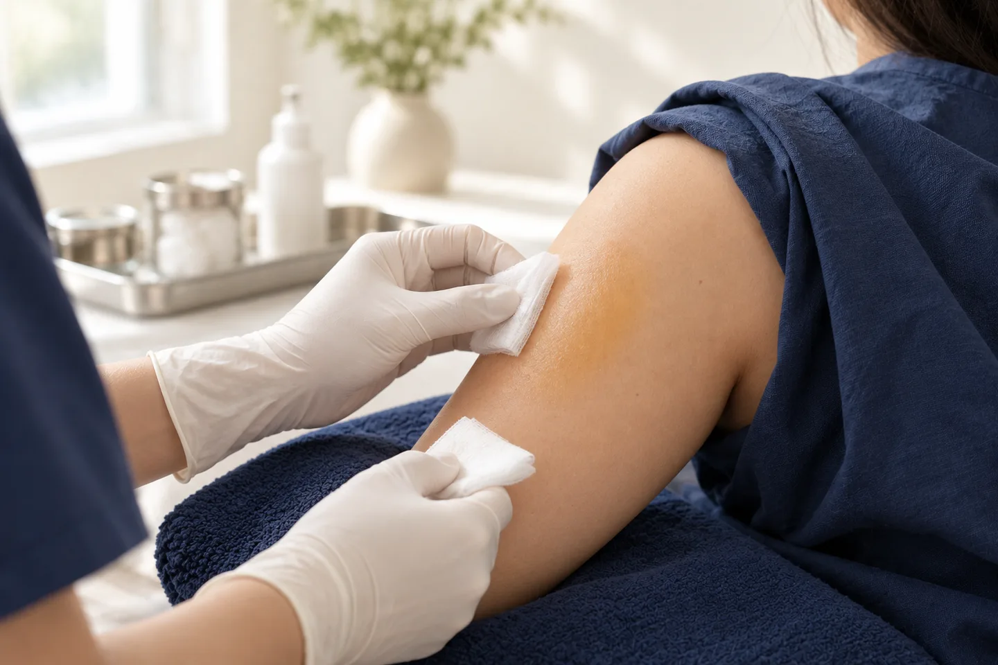
For a typical small superficial lipoma, removal is often performed as Minor Skin Surgery using local anesthesia. The dermatologist makes an incision over the lump, carefully separates the lipoma from the surrounding tissue, removes it, controls bleeding, and closes the wound with sutures when appropriate.
The most important step, however, happens before surgery: confirming that the lump behaves like a benign lipoma. A rapidly growing, firm, fixed, deep, painful, or unusually large soft-tissue mass may need imaging, biopsy, or specialist evaluation before excision rather than routine minor surgery.
What Is a Lipoma?
A lipoma is a benign tumor composed predominantly of mature adipocytes, or fat cells. It commonly occurs within the subcutaneous tissue between the skin and deeper structures.
Typical lipomas tend to be:
- Soft or rubbery
- Smooth
- Mobile under the skin
- Slow-growing
- Usually painless
- Covered by normal-looking skin
Common locations include the:
- Neck
- Shoulders
- Upper back
- Arms
- Abdomen
- Thighs
Some people develop only one lipoma, while others develop several.
Is a Lipoma Cancer?
A typical lipoma is benign and is not cancer.
This distinction is important because not every fatty or soft-tissue mass is necessarily a lipoma.
A malignant tumor of fatty tissue, such as Liposarcoma, is a different condition. A suspicious soft-tissue mass should not simply be assumed to be a lipoma and removed without appropriate assessment.
The purpose of a dermatology assessment is therefore not simply to decide whether a lump can be removed, but first to answer:
What is this lump, and is routine Lipoma Excision appropriate?
What Causes Lipomas?
The exact reason an individual lipoma develops is often unclear.
Lipomas are growths of mature fat cells rather than simply collections of excess body fat. They can therefore occur in people of different body sizes.
Some people appear to have a tendency to develop multiple lipomas, and multiple lipomas can occasionally be associated with inherited conditions or syndromes.
Having a lipoma does not necessarily indicate an unhealthy diet or excess body fat.
What Does a Lipoma Feel Like?
A classic superficial lipoma usually feels like a soft or doughy lump underneath the skin.
When gently pressed, it may move slightly rather than feeling attached to the skin or deeper tissue.
The skin over it usually appears normal.
Many lipomas remain asymptomatic for years. Others become noticeable because they:
- Increase in size
- Rub against clothing
- Become uncomfortable with movement
- Cause pressure symptoms
- Become painful
- Create a visible contour
- Become cosmetically bothersome
A lump that does not behave like a typical lipoma deserves closer evaluation.
Can a Lipoma Be Painful?
Yes, although many lipomas are painless.
Pain or tenderness may occur because of the location of the lesion, pressure on surrounding structures, trauma, or a particular lipoma variant.
However, pain is also one of the features that may lead a clinician to investigate a soft-tissue mass more carefully, particularly if it is accompanied by rapid growth, firmness, fixation, or unusual depth.
When Should a Lipoma Be Removed?
Not every lipoma needs surgery.
Observation is often reasonable when a lump has typical benign features, remains stable, causes no symptoms, and the diagnosis is clinically reassuring.
Lipoma Excision may be considered when the lesion:
- Is painful or tender
- Is progressively enlarging
- Causes pressure or discomfort
- Interferes with movement
- Frequently rubs against clothing
- Is cosmetically bothersome
- Creates a noticeable contour
- Causes diagnostic uncertainty
- Needs Histopathology for confirmation
Surgical removal is the standard definitive treatment for many symptomatic superficial lipomas.
When Should a Lump Be Investigated Before Excision?
This is one of the most important questions for anyone considering Lipoma Excision.
Most superficial soft-tissue lumps are benign, but some features require further assessment.
A lump deserves particular attention if it is:
- Growing rapidly
- Approximately 5 cm or larger
- Located deep to the fascia
- Firm or hard rather than soft
- Fixed to deeper tissue
- Painful without an obvious explanation
- Suddenly enlarging
- Recurrent after previous removal
- Associated with neurological symptoms
- Located in an anatomically complex area
- Clinically atypical for a lipoma
Soft-tissue masses that are deep, rapidly growing, or 5 cm or larger warrant more careful evaluation because these features can occur in soft-tissue malignancies.
Importantly, size alone does not mean that a lump is cancerous. Large lipomas certainly occur. Size is a reason for additional assessment, not a diagnosis.
Lipoma vs Liposarcoma: What Is the Difference?
Patients frequently worry that a lump described as fatty could be Liposarcoma.
They are different conditions.
| Lipoma | Concerning Soft-Tissue Mass |
|---|---|
| Usually benign | Requires evaluation to exclude malignancy |
| Often slow-growing | Rapid growth is concerning |
| Usually soft | May be firm |
| Often mobile | Fixation may be concerning |
| Usually superficial | Deep location deserves assessment |
| Often painless | Unexplained pain can require investigation |
| Often clinically recognizable | May require imaging and/or biopsy |
This table is useful for understanding warning signs, but it cannot be used to diagnose a lump at home.
Imaging or biopsy may be required when a suspected lipoma is large, unusual, or appears deeper than expected.
How Does a Dermatologist Diagnose a Lipoma?
Diagnosis begins with history and physical examination.
The dermatologist may ask:
- When did you first notice the lump?
- Has it increased in size?
- How quickly is it growing?
- Is it painful?
- Has it changed recently?
- Have you had similar lumps?
- Has the area been treated previously?
- Do family members have multiple lipomas?
During examination, the clinician assesses:
- Size
- Location
- Consistency
- Mobility
- Depth
- Relationship to the skin
- Relationship to deeper structures
- Tenderness
- Surface changes
Many typical superficial lipomas can be recognized clinically without extensive testing.
Does Every Lipoma Need an Ultrasound?
No.
A small, superficial, freely mobile lump with classic features of a lipoma may not require imaging before removal.
An Ultrasound can be useful when the diagnosis or anatomy is less clear. It can help assess:
- Size
- Depth
- Whether the lesion is solid or cystic
- Relationship to surrounding tissue
- Vascularity
For deeper or more concerning masses, additional imaging such as MRI may be more appropriate.
When Is MRI Needed for a Suspected Lipoma?
MRI may be considered when a mass is:
- Deep
- Large
- Rapidly growing
- Fixed
- Painful
- Located near important anatomical structures
- Clinically atypical
- Difficult to characterize with examination or Ultrasound
MRI provides substantially more information about the relationship between a soft-tissue mass and muscles, fascia, nerves, and other deeper structures.
A suspicious deep soft-tissue mass should be evaluated appropriately before attempting routine excision.
Lipoma vs Epidermoid Cyst
These are commonly confused because both can present as lumps beneath the skin.
Lipoma
A lipoma is composed of fatty tissue and is usually soft, mobile, and located beneath the skin.
Epidermoid Cyst
An Epidermoid Cyst is a sac-like lesion containing keratin. It may be more closely attached to the skin and sometimes has a visible central punctum.
Epidermoid Cysts can become acutely inflamed, red, swollen, and painful.
Treatment is also different: removing an Epidermoid Cyst requires removal of its cyst wall, whereas Lipoma Excision involves separating and removing fatty tumor tissue.
Lipoma vs Other Lumps Under the Skin
Other conditions that may resemble a lipoma include:
- Epidermoid Cyst
- Dermatofibroma
- Abscess
- Enlarged lymph node
- Hematoma
- Vascular lesion
- Peripheral nerve tumor
- Other benign soft-tissue tumors
- Soft Tissue Sarcoma
- Liposarcoma
This is why a persistent lump should ideally be diagnosed before deciding on treatment.
What Is Lipoma Excision?
Lipoma Excision is a surgical procedure in which the lipoma is removed through an incision in the skin.
For an appropriate superficial lipoma, this is usually considered Minor Skin Surgery.
The goal is to remove the lipoma while protecting surrounding structures and creating a wound that can heal with an acceptable scar.
Complete surgical removal is generally preferred when definitive treatment is desired.
How Is Lipoma Excision Performed?
The exact technique depends on the size, location, depth, and anatomy of the lesion.
A typical procedure may involve the following steps.
1. Clinical Assessment
The dermatologist confirms that the lesion appears suitable for minor surgical removal.
If the mass is atypical, further investigation may be recommended before surgery.
2. Marking the Lipoma
The borders and planned incision may be marked while the lump is easily palpable.
3. Cleaning the Skin
The surgical area is cleaned using an appropriate antiseptic technique.
4. Local Anesthesia
Local anesthetic is injected around the surgical area to numb the skin and deeper tissues.
The injection may sting briefly.
Once adequate anesthesia has taken effect, the patient should generally feel pressure or movement rather than sharp pain.
5. Skin Incision
A controlled incision is made over or adjacent to the lipoma.
The length and direction of the incision depend on several factors, including the size and location of the lesion and the need to remove it safely.
6. Dissection
The lipoma is carefully separated from surrounding tissue.
Many superficial lipomas are relatively well circumscribed, but not every lesion has a simple capsule that allows it to “pop out.
”
Some lesions may extend between tissue planes or deeper into surrounding structures.
7. Removal
The lipoma is removed, ideally completely when clinically appropriate.
8. Hemostasis
Bleeding is controlled before the wound is closed.
9. Suturing
The wound may be closed in one or more layers depending on its depth.
Deeper wounds may require buried absorbable sutures as well as superficial sutures.
10. Dressing
A dressing is applied and wound-care instructions are provided.
Is Lipoma Excision Performed Under Local Anesthesia?
Many small and uncomplicated superficial lipomas can be removed using local anesthesia.
This means the patient remains awake while the surgical area is numb.
However, local anesthesia is not necessarily suitable for every lipoma.
Very large, deep, intramuscular, anatomically complex, or otherwise difficult lesions may require treatment in a different surgical setting or referral to an appropriate specialist.
The safest setting should be determined by the characteristics of the mass rather than simply by patient preference.
Does Lipoma Excision Hurt?
The local anesthetic injection can cause temporary stinging or burning.
During the procedure, patients may notice:
- Pressure
- Pulling
- Movement
- Touch
They should not normally experience sharp surgical pain once adequate anesthesia has been achieved.
Some soreness after the anesthetic wears off is expected.
The degree of discomfort depends on the size, location, depth, and extent of surgery.
How Long Does Lipoma Excision Take?
There is no single procedure time for every lipoma.
A small, superficial, easily accessible lesion may be relatively straightforward.
A larger or deeper lesion may require substantially more time because the surgeon must:
- Dissect a larger area
- Identify tissue planes
- Control bleeding
- Protect nearby structures
- Close a deeper surgical defect
For this reason, examination before scheduling surgery can be important.
Does a Lipoma Need to Be Removed in One Piece?
When practical, complete removal of the lesion is generally desirable.
However, surgical technique should be determined by anatomy and safety rather than by the goal of producing an intact specimen at all costs.
The surgeon must avoid unnecessary injury to surrounding structures.
What Happens to the Lipoma After Removal?
The excised tissue may be submitted for Histopathology, where a pathologist examines the tissue microscopically.
Histopathology can:
- Confirm that the lesion is a lipoma
- Identify a lipoma subtype
- Detect unexpected pathology
- Help clarify an uncertain diagnosis
A typical lipoma microscopically consists predominantly of mature adipocytes.
Whether Histopathology is recommended routinely can depend on the clinical context and local practice, but it is particularly valuable when the lesion is atypical or the diagnosis is uncertain.
Can Lipoma Excision Be Done Through a Very Small Incision?
Various minimal-incision techniques have been described for selected lipomas, and smaller incisions may sometimes produce shorter scars, reducing overall scarring.
However:
The smallest possible incision is not always the safest or best incision.
A very small opening can make visualization, bleeding control, complete removal, and protection of surrounding structures more difficult.
The incision should be appropriate for the lipoma’s:
- Size
- Depth
- Location
- Mobility
- Anatomy
The goal should be safe removal with thoughtful scar placement, not simply the shortest incision.
Will Lipoma Excision Leave a Scar?
Yes.
Any procedure that requires a skin incision produces a scar.
The final appearance varies according to:
- Size of the lipoma
- Incision length
- Location
- Wound tension
- Individual healing tendency
- Infection or wound complications
- Sun exposure
- History of Keloid or Hypertrophic Scar
- Postoperative wound care
Initially, a scar may appear pink, red, brown, firm, or raised.
Scar maturation takes time and can continue for months.
Can a Lipoma Be Removed Without a Scar?
No method that removes a subcutaneous lipoma through the skin can guarantee a scar-free result.
Claims of “scarless Lipoma Removal” should therefore be interpreted cautiously.
Even very small incisions can leave visible scars.
Lipoma Excision and Asian Skin
Lipoma Excision can be performed in Asian skin, but postoperative scar appearance deserves particular attention.
Patients with more pigmented skin may develop:
- Post-Inflammatory Hyperpigmentation
- Temporary darker coloration around the scar
- Hypopigmentation
- Hypertrophic Scar
- Keloid in predisposed individuals
A personal history of Keloid or unusually thick scars should be discussed before surgery.
Can Lipoma Be Removed With Liposuction?
Liposuction has been used to reduce or remove selected lipomas and may involve smaller access sites as one treatment option.
However, it has limitations.
Compared with conventional excision, it may:
- Make complete removal more difficult
- Fragment the specimen
- Make pathological assessment less straightforward
- Be unsuitable when the diagnosis is uncertain
For many discrete superficial lipomas requiring definitive removal, surgical excision remains a straightforward approach.
Can Lipomas Be Treated With Laser?
A typical subcutaneous lipoma is fundamentally different from a superficial epidermal lesion such as a Skin Tag or Seborrheic Keratosis.
Because the lesion is located beneath the skin, simply treating the surface with a laser does not remove the underlying fatty mass.
Any proposed technique should be evaluated based on whether it can safely and adequately address the actual subcutaneous lesion.
Can Lipomas Be Removed With Injections?
Injection-based approaches have been explored for selected fatty lesions, but they are not equivalent to complete surgical excision.
When the diagnosis is uncertain, destroying or altering a mass without obtaining appropriate tissue can also complicate diagnostic assessment.
Patients seeking definitive treatment should discuss the advantages and limitations of each method before proceeding.
What Are the Risks of Lipoma Excision?
Lipoma Excision is generally a minor surgical procedure for appropriately selected superficial lesions, but it is still surgery.
Possible complications include:
- Pain
- Bleeding
- Bruising
- Swelling
- Hematoma
- Infection
- Wound separation
- Delayed healing
- Fluid collection or Seroma
- Scar
- Post-Inflammatory Hyperpigmentation
- Hypertrophic Scar
- Keloid
- Temporary or persistent numbness
- Nerve injury
- Injury to surrounding structures
- Incomplete removal
- Recurrence
The risk profile varies considerably according to location, size and depth. Deep or anatomically complex masses may carry risks that are very different from those of a small superficial lipoma.
Can Lipoma Excision Cause Numbness?
Yes.
Small sensory nerves run through subcutaneous tissue and may be stretched, irritated, or injured during surgery.
Temporary numbness around a surgical scar can occur.
The risk of significant nerve injury depends heavily on anatomy. A mass close to an important nerve requires more careful surgical planning and may not be appropriate for routine office-based Minor Skin Surgery.
Can a Lipoma Come Back After Excision?
Recurrence after complete removal of a typical lipoma is generally uncommon, but it is possible.
A lump appearing again at the same location may represent:
- Residual lipoma
- Recurrence
- Scar tissue
- Another benign lesion
- A different soft-tissue mass
A recurrent mass should be reassessed rather than automatically undergoing repeat removal.
Can I Develop Another Lipoma Somewhere Else?
Yes.
Removing one lipoma does not prevent another independent lipoma from developing elsewhere.
This is particularly relevant for people who naturally develop multiple lipomas.
Recovery After Lipoma Excision
Recovery varies according to the extent of surgery.
A small superficial lipoma may leave a relatively small wound, whereas removal of a large mass can create a significant space beneath the skin.
Immediately after surgery, patients may experience:
- Mild pain
- Tenderness
- Swelling
- Bruising
- Tightness around the sutures
These symptoms generally need to be interpreted in relation to the size and location of the operation.
Wound Care After Lipoma Excision
Patients should follow the specific instructions provided by their treating clinician.
General principles may include:
- Keep the initial dressing clean and dry as instructed
- Wash the wound according to the postoperative plan
- Avoid picking at crusts or sutures
- Avoid unnecessary friction
- Avoid excessive stretching of the wound
- Attend scheduled wound checks
- Return for suture removal when required
- Watch for signs of infection
Instructions may differ depending on whether the wound is on the face, trunk, arm, leg, scalp, or another location.
When Are Sutures Removed?
Suture-removal timing depends primarily on anatomical location, wound tension, and the type of closure.
There is no single correct day for every Lipoma Excision.
Some buried sutures are absorbable and do not need removal, while external sutures usually need to be removed at an appropriate follow-up visit.
Patients traveling internationally should discuss this before surgery.
When Can I Exercise After Lipoma Excision?
This depends on the location and size of the wound.
Exercise places tension on surgical wounds and can increase:
- Bleeding
- Swelling
- Wound separation
- Scar widening
A small wound in a low-tension area may require relatively little restriction, whereas a larger wound over the shoulder, back, arm, thigh, or near a joint may require a longer period of activity modification.
Ask the treating clinician specifically about:
- Gym training
- Running
- Swimming
- Yoga
- Contact sports
- Heavy lifting
When Can I Swim After Lipoma Excision?
Swimming should generally wait until the wound has sealed adequately and the clinician considers it safe.
Swimming too early may expose a healing wound to moisture, contamination, friction, and prolonged soaking.
This is particularly relevant for patients visiting Bangkok who plan to travel to beaches or islands elsewhere in Thailand shortly after surgery.
Can I Fly After Lipoma Excision?
Flying itself is not automatically prohibited after minor superficial Lipoma Excision.
The more important considerations are:
- Wound size
- Bleeding risk
- Pain
- Need for dressing changes
- Suture removal
- Histopathology results
- Access to medical care if a complication occurs
International patients should ideally avoid arranging surgery immediately before a departure that makes appropriate follow-up impossible.
Warning Signs After Lipoma Excision
Contact your treating clinician if you develop:
- Increasing rather than improving pain
- Progressive redness
- Increasing warmth
- Pus or abnormal discharge
- Fever
- Significant swelling
- Persistent bleeding
- Wound separation
- Rapidly enlarging swelling beneath the wound
- New significant numbness or weakness
- Skin discoloration suggesting compromised blood supply
Some swelling, bruising, and discomfort can be normal after surgery. The key concern is symptoms that are severe, progressive, or unexpected.
When Should You See a Dermatologist for a Lump?
Consider a medical assessment if you discover a new persistent lump, particularly when you are unsure what it is.
Seek more prompt evaluation when the lump:
- Grows rapidly
- Is approximately 5 cm or larger
- Feels deep
- Is fixed
- Is unusually firm
- Becomes increasingly painful
- Recurs after removal
- Causes neurological symptoms
- Has an atypical appearance
These features do not automatically mean cancer, but they are reasons not to assume that the lesion is a simple lipoma.
What to Expect at a Lipoma Consultation in Bangkok
A useful consultation should answer more than “Can this lump be removed?”
The clinician should first establish:
Is this actually a lipoma?
A clinical examination may be sufficient for a typical superficial lesion. An atypical mass may require imaging or further investigation.
How deep is it?
Depth affects both diagnosis and surgical planning.
Is Minor Skin Surgery appropriate?
A small superficial lipoma and a large deep intramuscular mass should not be managed as if they were the same procedure.
Is imaging needed?
Ultrasound or MRI may be considered according to the clinical findings.
What scar should I expect?
The likely incision should be discussed before surgery, particularly when the lesion is in a cosmetically sensitive location.
Will the tissue be sent for Histopathology?
This should be clarified before the procedure.
What follow-up is required?
Patients should understand wound care, suture removal, activity restrictions, pathology follow-up, and warning signs before leaving.
Lipoma Excision for Tourists and International Patients in Bangkok
Bangkok is a common destination for international medical care, but Minor Skin Surgery should still be planned around safe postoperative follow-up.
If you are visiting Thailand for a limited period, consider:
- Whether consultation and surgery can occur on the same trip
- Whether imaging is needed before surgery
- How long you should remain in Bangkok after the procedure
- When sutures need removal
- Whether you can continue wound care while traveling
- Whether swimming should be postponed
- When Histopathology results will be available
- How results will be communicated after you leave Thailand
- Where to seek medical attention if the wound becomes infected
- Whether planned flights or strenuous activities interfere with recovery
For a traveler, the “fastest” surgical schedule is not necessarily the safest one.
Frequently asked questions
Is Lipoma Excision a major operation?
A small superficial lipoma can often be treated as Minor Skin Surgery. Large, deep, intramuscular, or anatomically complex lesions may require a different surgical setting.
Does every lipoma need removal?
No. Typical asymptomatic lipomas can often be observed.
When should a lipoma be removed?
Removal may be considered for pain, growth, discomfort, cosmetic concerns, functional problems, or diagnostic uncertainty.
Is a lipoma cancer?
A typical lipoma is benign. However, not every soft-tissue lump is necessarily a lipoma.
Can a lipoma become Liposarcoma?
Lipoma and Liposarcoma are distinct tumors. The practical concern is that an atypical mass initially assumed to be a lipoma may require further investigation to establish the correct diagnosis.
What are the warning signs that a lump may not be a simple lipoma?
Rapid growth, deep location, fixation, significant size, unexplained pain, or atypical examination findings warrant further assessment.
Does a lipoma need Ultrasound before removal?
Not always. Typical small superficial lipomas may be diagnosed clinically. Ultrasound can be useful when size, depth, or diagnosis is uncertain.
Does a lipoma need MRI?
Not routinely. MRI is more relevant for deep, large, atypical, or concerning soft-tissue masses.
Is Lipoma Excision painful?
Local anesthetic injection can sting briefly. Once adequately numb, sharp surgical pain should generally not be felt.
Can Lipoma Excision be done under local anesthesia?
Many small superficial lipomas can be removed under local anesthesia. Larger or deeper lesions may require a different approach.
How large will the incision be?
It depends on the size, location, depth, mobility, and surgical technique. A safe incision is more important than simply making the smallest possible incision.
Will Lipoma Excision leave a scar?
Yes. Any surgical incision leaves a scar, although its final appearance varies considerably.
Can a lipoma be removed without a scar?
No technique can guarantee scar-free removal.
Can a lipoma be removed with Laser?
A typical lipoma is a subcutaneous fatty mass, so treating only the skin surface does not remove the underlying lesion.
Can Liposuction remove a lipoma?
It may be used in selected cases, but it has limitations compared with conventional surgical excision.
Should a removed lipoma be sent for Histopathology?
Histopathology can confirm the diagnosis and is particularly important when the clinical diagnosis is uncertain or the lesion has atypical features.
Can a lipoma return after removal?
Recurrence is possible, although it is generally uncommon after complete removal of a typical lipoma.
Can another lipoma develop elsewhere?
Yes. Removing one lipoma does not prevent new independent lipomas.
How long does recovery take?
Recovery depends on size, depth, location, wound tension, and the type of closure.
When can I return to work?
Many patients can return relatively quickly after minor surgery, but jobs involving heavy lifting or repetitive movement may require longer activity modification.
When can I exercise?
This depends on wound location and size. Exercise that pulls on the wound may need to be temporarily restricted.
When can I swim?
Wait until the wound has adequately healed and your clinician considers swimming safe.
Can I fly after Lipoma Excision?
Often yes after uncomplicated minor surgery, but follow-up, wound care, suture removal, and Histopathology should be planned before traveling.
What happens if my lump grows rapidly?
A rapidly growing mass should be reassessed rather than simply booked for routine Lipoma Excision.
Should a recurrent lipoma be removed again?
It should first be reassessed to confirm what the recurrent lump represents.
Related conditions
-
Lipomas Lipomas A lipoma is a benign tumor made of mature fat cells. It usually develops slowly beneath the skin and typically feels soft, rubbery, and movable when pressed. Lipomas commonly occur on the trunk, shoulders, neck, arms, back, abdomen, and thighs. They are not cancer and often do not require treatment.
-
Cysts Cysts A skin cyst is a lump that develops beneath or within the skin. One of the most common types seen in dermatology is the Epidermoid Cyst, sometimes incorrectly called a “sebaceous cyst.

-
Cyst Excision Cyst Excision A skin cyst is a lump that develops beneath or within the skin. One of the most common types seen in dermatology is the Epidermoid Cyst, sometimes incorrectly called a “sebaceous cyst.

-
Skin Biopsy (การวินิจฉัย / Diagnosis) Skin Biopsy (การวินิจฉัย / Diagnosis) A skin biopsy is a minor medical procedure in which a dermatologist removes a small sample of skin so it can be examined under a microscope. It is one of the most important diagnostic tools in dermatology because many skin conditions cannot be confirmed by appearance alone.